Article
The Biophotonics Imaging Lab at the Beckman Institute for Advanced Science and Technology has developed imaging techniques that investigate tissues without using any staining or labels. The researchers created a unique system using a laser source that can capture more information about a tissue compared to traditional imaging techniques. That system provides better visualization of extracellular vesicles — small packages which are known to increase in number and be associated with cancer — particularly in connection to breast cancer cells.
“We can acquire information about the structure and metabolism of the living tissue. The imaging system allows us to capture all this information simultaneously, allowing us to see many more details about the tissue, cells, and their functions than the current ways of imaging,” said Dr. Stephen Boppart, the Abel Bliss Professor of Engineering with appointments in electrical and computer engineering and bioengineering at the University of Illinois at Urbana-Champaign, who leads the Biophotonics Imaging Lab, and is also a medical doctor.
The paper “Label-Free Visualization and Characterization of Extracellular Vesicles in Breast Cancer” was published in the Proceedings of the National Academy of Sciences, and highlighted on the cover of the journal.
“Cells use these extracellular vesicles to communicate with each other, even under normal conditions. Tumor cells will alter these extracellular vesicles, and release more throughout the body. For this reason, they have the potential to be used as markers for cancer progression,” Boppart said.
Traditional visualization techniques are dependent on the use of labels, which involves a lot of effort and can render the tissue unusable for additional investigation. “Our key contribution is that we can visualize these vesicles in living tissues without perturbing the tissues or using any labels. We can see how these vesicles interact with each other and with the tumors,” said Sixian You, a bioengineering graduate research assistant in the Boppart lab who is first author on the paper.
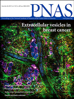 The study, “Label-Free Visualization and Characterization of Extracellular Vesicles in Breast Cancer,” was published in the Proceedings of the National Academy of Sciences in November 2019 and highlighted on the journal’s cover.
The technique used by the lab involves using ultrashort laser pulses which interact with the tissue samples.
“There are two mechanisms involved in imaging," You said. "Some of the tissue components emit different kinds of fluorescence which comes in different colors. The other mechanism involves molecular structures that when aligned in a certain way, will give you an entirely different set of colors.”
The researchers have also begun to characterize the contents of the vesicles using this technique.
“We have found that the vesicles which have higher concentrations of NADPH molecules are highly correlated with tumor aggressiveness, and are often located around vessels. By characterizing the vesicles, we have a good shot at detecting cancerous changes at an earlier stage,” You said.
A second paper “Real-Time Intraoperative Diagnosis by Deep Neural Network Driven Multiphoton Virtual Histology” was published in Precision Oncology, and it looks at combining the label-free imaging with deep learning techniques.
Currently, when surgeons remove tumors from patients, they analyze the tissue samples from the margins of the tumor to ensure that all the cancer tissue has been removed. Unfortunately, the pathology labs that analyze the samples can take up to a few days to complete their analysis.
“We are trying to use the label-free technique to look at the tissue right away in the surgical room,” You said. “After we get the images we use deep learning, which can be used to differentiate between cancerous and normal breast tissue.”
“We hope that this technique will be used for medical diagnosis and more clinical applications. The current techniques are useful but outdated and time-consuming. We think that there is a lot of new information that we can gain from this technology,” Boppart said.
“The system itself is somewhat expensive, but not unlike other high-end microscopes currently in use, and costs should come down in a few years as the laser technology matures,” Boppart said. “The other inherent challenge for us is that if you want high resolution images, you need to focus on smaller areas. However, when we look at the smaller areas, we can still get an adequate picture of what is going on in the tissue.”
Boppart recently discussed the work on the Future Tech Podcast.
The paper “Label-Free Visualization and Characterization of Extracellular Vesicles in Breast Cancer” is online at doi.org/10.1073/pnas.1909243116
The paper “Real-Time Intraoperative Diagnosis by Deep Neural Network Driven Multiphoton Virtual Histology” can be found at https://www.nature.com/articles/s41698-019-0104-3
Beckman Institute for Advanced Science and Technology