Research images
Science is beautiful.
Every day, researchers use the Beckman Institute's core facilities to conduct barrier-breaking research. They also create stunning images along the way. Click on each image to learn more.
Follow @beckmaninstitute on Instagram for the latest.
Beckman Institute Research Image Contest
Each spring, Beckman hosts a research image contest that includes categories for undergraduates, graduates, postdoctoral or staff researchers, and faculty members.
Members of the Beckman community are invited to enter, as are those who work outside Beckman but have used the institute's core facilities, the Biomedical Imaging Center, Microscopy Suite, Molecular Imaging Laboratory and Visualization Laboratory, to capture images.
Leaders of those facilities and the Beckman director serve as blind judges. Winning images are framed and displayed for one year in the Beckman Atrium, then another in the Director's Conference Room. Then, they are displayed on the walls around the institute and throughout the community.
Beckman researchers should watch their email and the Beckman Bulletin each spring for information about the contest. Questions can be directed to the Communications Office.
Research image news
Six framed images showing (and explaining) a more intimate view of research at the Beckman Institute are being featured in the institute’s director’s conference room.
The art was selected through an internal contest that challenged undergraduate and graduate students, and postdoctoral researchers to submit images showing the intersection between art and science. Each category features a winner and honorable mention.
“At the Beckman Institute, we celebrate the collaborative and interdisciplinary research happening within our walls,” said Jeff Moore, director of the Beckman Institute and an Ikenberry Endowed Chair in the Department of Chemistry. “I’m excited about showcasing images captured using our core research facilities, by the students and postdocs doing incredible work in this space.”
The director’s conference room previously showcased the portraits of founder Arnold Beckman and all of the institute’s previous directors. Those portraits have been moved to prominent locations near the Beckman Institute’s third-, fourth-, and fifth-floor tower rooms.
The first-place winners and honorable mention recipients, respectively, and a description of their image:
Undergraduate Students
Elizabeth Murphy, chemistry, Braun Research Group
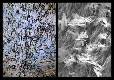
Proteins have proven to have tremendous potential as biological pharmaceuticals and drug targets. The key to successfully using proteins for these applications is maintaining stability. There is widespread interest in the use of zwitterionic poly(sulfobetaine) (pSB) to stabilize proteins in solution due to their assumed “protein-avoidance.” However, pSB-protein interactions have yet to be completely understood. For example, despite the existence of complementary charged pSB and proteins, why would pSB not interact with proteins? This research demonstrated that pSB chains in solution can actually interact with proteins directly and affect their stability. Displayed is an inspection light microscope image of pSB in solution.
Andrey Nosatov, materials science and engineering, Harley Research Lab
Crystals within mineralized collagen scaffolds used in composite materials to create bone replacements. The crystals were created by lyophilizing a mix of collagen and calcium- and phosphate-containing compounds to lead to mineralization. This image was captured on an environmental scanning electron microscope.
Graduate Students
Jaena Park, second-year Ph.D. student in bioengineering, Biophotonics Imaging Laboratory and Mayo Clinic
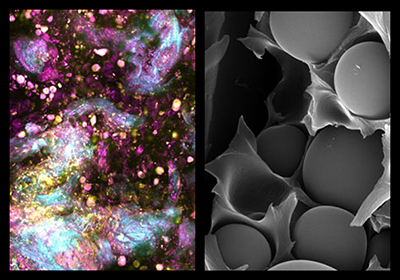
An image of a human pancreatic adenocarcinoma growing in a living avatar or mouse, taken by simultaneous label-free autofluorescence multi-harmonic microscopy. Pink and yellow objects are cells in the tumor tissue, and collagen is blue. Our research uses these images to characterize extracellular vesicles formed in the tumor.
Kelly Chang, third-year Ph.D. student in materials science and engineering, Autonomous Materials Research Group
This is a micrograph of epoxy microbeads embedded in a thermoplastic-rich region of a self-healing material. By combining thermoplastic with epoxy, we are able to make tougher composites. To add self-healing capabilities, we can also add liquid-filled microcapsules. When a material containing these microcapsules is damaged, the liquid releases from the capsules, reacts with the thermoplastic, and initiates self-healing. In particular, this system of epoxy mixed with thermoplastic and microcapsules are incorporated in carbon fiber composites for aerospace applications (think tough self-healing airplanes!).
Postdoctoral Researchers
Sudipta Mukherjee, Chemical Imaging and Structures Laboratory
Research area: chemical imaging
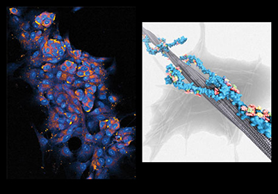
This stimulated Raman scattering microscopy image shows a co-culture of colon cancer cells and fibroblasts embedded in hydrogel matrix. SRSM is a label-free technique that relies on chemical specificity of Raman active vibrational modes to generate contrast. The blue represents the nuclei and the yellow spots are from lipid droplets. One or more distinct nucleoli inside the nuclei are also visible. The image is of a model sample wherein we try to simulate the tumor microenvironment in an effort to understand the signaling between fibroblasts and cancer cells and its consequence in the growth and progression of cancer. Image collected by Sudipta Mukherjee and Sanghamitra Deb.
Giuseppe Licari, Theoretical and Computational Biophysics Group
Research area: macromolecules interaction and molecular recognition
A molecular model of a novel compound wrapping around a β-amyloid fibril. This compound, a multivalent polymer-peptide conjugate (see the polymer backbone in cyan and active peptides in multicolor), inhibits the formation of the β-amyloid fibril, which plays a fundamental role in the development of Alzheimer’s disease. A transmission electron microscope image in the background illustrates in vitro fibril aggregation. Our research is trying to understand the mechanism of amyloid aggregation and develop new molecular architectures capable of inhibiting such phenomenon.
Beckman Institute for Advanced Science and Technology