Article
What do a synthetic chemist, a medical imaging expert, and a neurologist have in common? They’re coming together in Beckman's Biomedical Imaging Center and Molecular Imaging Laboratory to develop better diagnostic tools and imaging agents to detect early-stage Alzheimer’s disease and other neurodegenerative diseases.
The dream team
-alongside-liviu-mirica-(center)-and-dr.-daniel-llano-.jpg?sfvrsn=ef50eef8_1) Wawryzneic “Wawosz” Dobrucki (left), alongside Liviu Mirica (center), and Dr. Daniel Llano.
Wawryzneic “Wawosz” Dobrucki (left), alongside Liviu Mirica (center), and Dr. Daniel Llano.
A team led by Liviu M. Mirica along with Wawrzyniec “Wawosz” Dobrucki and Dr. Daniel A. Llano received a $3 million grant from the U.S. National Institute on Aging of the National Institutes of Health to develop and test multi-modal imaging agents for the detection of Alzheimer’s disease and related dementias. This grant is one of the first federal grants to bridge Beckman’s Magnetic Resonance Imaging Laboratory and Molecular Imaging Laboratory.
“I’m really excited about the opportunity to collaborate with different scientists from different fields,” said Mirica, a synthetic chemist and the William H. and Janet G. Lycan Professor of Chemistry in the School of Chemical Sciences at the University of Illinois Urbana-Champaign. His research group specializes in building and characterizing synthetic inorganic molecules in vitro: outside of the body.
Dobrucki, the Neil and Carol Ruzic Scholar for Biomedical and Translational Sciences, is an imaging expert who works extensively with PET scanning in Beckman’s Molecular Imaging Laboratory.
“I’m looking forward to high-resolution imaging of the brain and its structures,” Dobrucki said.
Llano, a professor of molecular and integrated physiology and a physician-surgeon, is a practicing neurologist who sees patients daily and specializes in in vivo brain studies: those inside the body.
“The potential impact that this project will have on Alzheimer’s is what I’m most excited about,” Llano said.
Understanding Alzheimer’s disease
Alzheimer’s disease is a neurodegenerative disease that negatively affects brain function and cognitive abilities. Along with Parkinson’s disease, amyotrophic lateral sclerosis, and other disorders, Alzheimer’s falls under the category of amyloid diseases. Amyloids are small groups of abnormally fibrous or misfolded proteins that do not commonly serve a purpose in the body.
A key marker of Alzheimer’s disease is the presence of amyloid plaques: large buildups of smaller beta-amyloid peptide aggregates. Peptides are short chains of amino acids that eventually create proteins. Neuroinflammation and oxidative stress in the brain are also major markers of Alzheimer’s.
The detection and treatment of neurodegenerative diseases is especially difficult because of the blood-brain barrier, a semipermeable system of blood vessels and capillaries that controls the flow of ions, molecules, and cells between the blood and the brain. To be effective, imaging agents and drug therapies (which are made of molecules or antibodies) need to be able to pass through.
Diagnosis and treatment
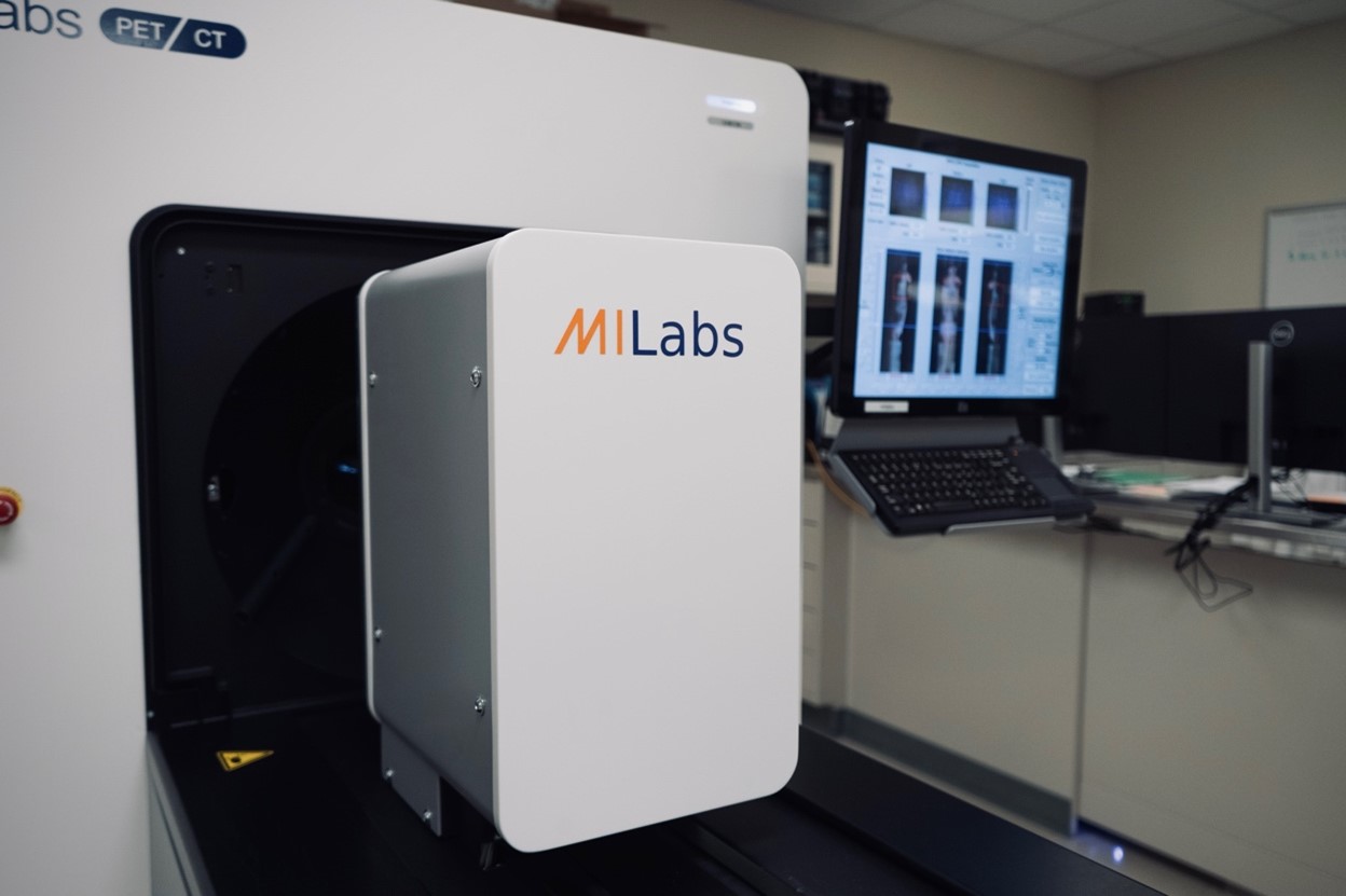
 This PET machine located in Beckman’s Molecular Imaging Laboratory will be operated by Dobrucki and used extensively during the team’s research.
Diagnosing Alzheimer’s disease with a high degree of accuracy requires identifying the amyloid aggregates and can only be completed during post-mortem investigation. This creates a need for diagnostic tools that can quickly locate soluble beta-amyloid peptide aggregates and larger amyloid plaques in a living patient.
PET and MRI are two noninvasive imaging methods commonly used in clinical settings. However, no MRI contrast agents that target amyloid aggregates have been developed. The few FDA-approved PET imaging agents are insufficient at detecting small-scale amyloid abnormalities or in some cases, lead to false-positives test results when diagnosing Alzheimer’s.
It's important to develop diagnostic tools to target smaller beta-amyloid peptides and other signs of neuroinflammation and oxidative stress for a variety of reasons, Mirica said. Creating multi-modal tools that can be used for both PET and MRI scans will give researchers a better idea of who is at risk for developing Alzheimer’s, who truly has the disease, and at what stage.
The $3M plan
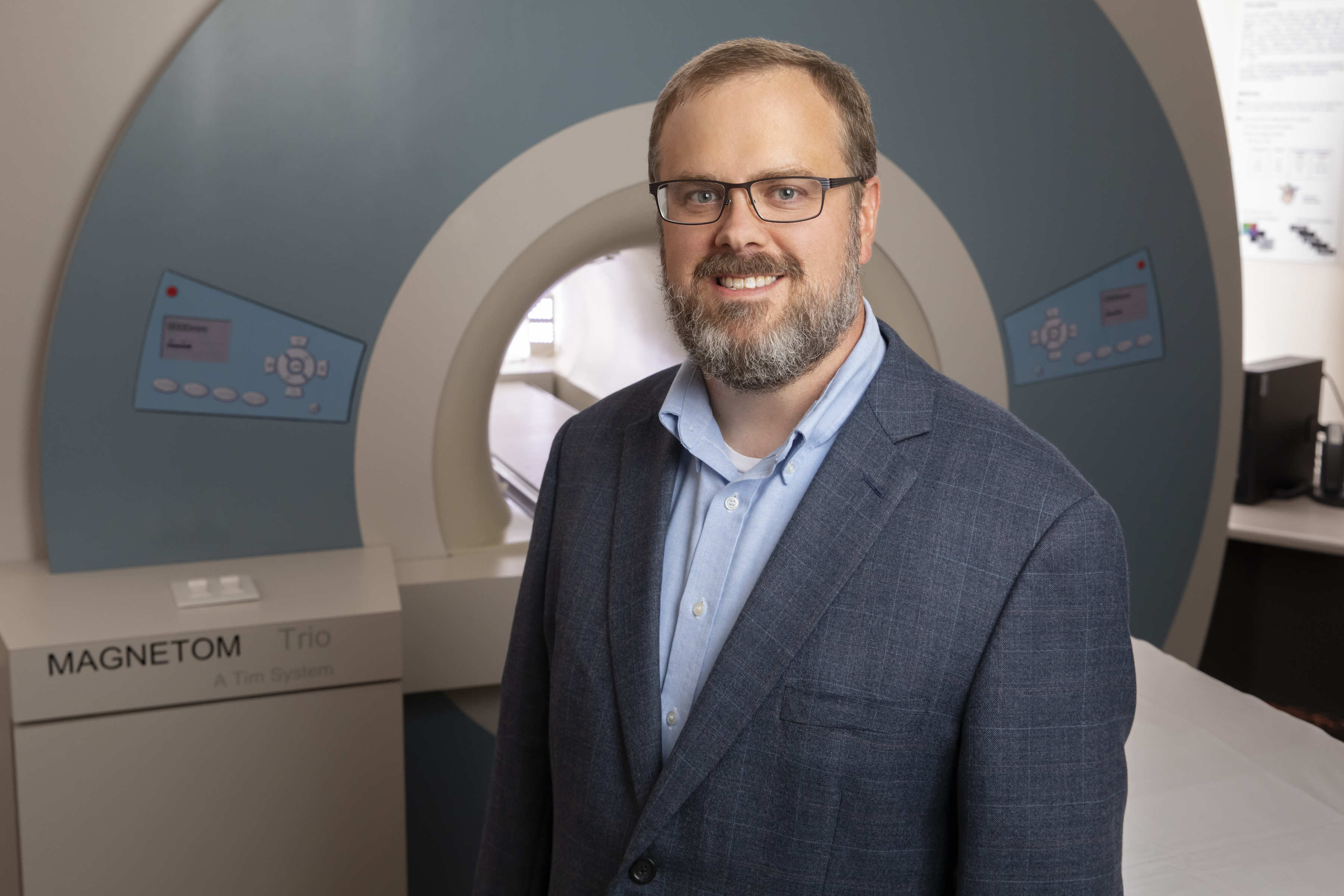
 Brad Sutton.
Mirica, Dobrucki, and Llano will receive the $3 million grant over the course of five years to generate novel dual-purpose imaging agents that can easily pass the blood-brain barrier and are compatible with both PET and MRI scanners.
This will enable the detection of neurodegenerative diseases at earlier stages and “will help tremendously in developing better therapies,” Mirica said.
Brad Sutton, a professor of bioengineering and the technical director of Beckman’s Biomedical Imaging Center, will assist the team by performing in vivo MRI studies. They will then evaluate the imaging agent’s ability as a dual modality diagnostic agent for Alzheimer’s disease and related dementias.
Already, Mirica and his collaborators have developed a series of customized molecules that can cross the blood-brain barrier and help detect both smaller soluble beta-amyloid peptides and larger insoluble amyloids.
They have also developed a copper-based PET imaging agent that led to the successful imaging of amyloid plaques in transgenic Alzheimer’s mice. Looking ahead, the team believes that these agents can be developed to pass through the blood-brain barrier in humans and image multiple markers of Alzheimer’s disease and other neurodegenerative diseases at earlier stages.
Editor's notes
Research reported in this press release was supported by National Institute on Aging of the National Institutes of Health under award number RF1AG083937. The content is solely the responsibility of the authors and does not necessarily represent the official views of the National Institutes of Health.
Mirica is also affiliated with the Carle Illinois College of Medicine and the Department of Bioengineering.
Dobrucki is also an associate professor of bioengineering and the associate head for graduate programs in the Department of Bioengineering.
Beckman Institute for Advanced Science and Technology