Article
When Sepideh Sadaghiani explains her work in cognitive neuroscience, she often quotes philosopher and psychologist William James. In The Principles of Psychology, James wrote: “Whilst part of what we perceive comes through our senses from the object before us, another part (and it may be the larger part) always comes out of our own mind.”
This idea that how we perceive the world has more to do with what is in our brain than on what our senses tell us was suggested by James more than 120 years ago. However, it’s only been in the last 20 years that neuroscientists have begun to be able to prove it.
Research, like Sadaghiani’s, that focuses on functional connectivity imaging is providing the evidence. In her lab at the Beckman Institute, Sadaghiani, an assistant professor of psychology and a member of the Intelligence, Learning, and Plasticity Group within the Intelligent Systems research theme, investigates cognitive control, network connectivity, and the relationship between the two in the brain.
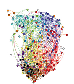
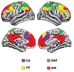
“Distant brain regions are in constant communication with each other,” she explained. “This communication, which is called functional connectivity, is foundational to all cognition. Functional connectivity is spatially organized into many large brain networks. But how this network organization is maintained and modulated in the service of flexible cognition, which is the mental ability to flexibly process internal and external information and goals, is poorly understood.”
Sadaghiani and her team are working to change that. She explained that until recently the assumption had been that the brain is “primarily a reactive machine and mainly fires in response to a stimulus or cognitive challenge, but we’re finding overwhelming evidence that most brain activity is in fact intrinsic and not dependent on external events. In fact, a growing body of studies, including ours, suggests that this intrinsic brain activity influences our perception of the world and cognition.”
To conduct those studies, Sadaghiani relies on functional magnetic resonance imaging (fMRI), a technique that uses MRI technology to measure brain activity by detecting changes associated with blood flow and electroencephalography (EEG), which permits direct observation of electrophysiological activity of the brain.
“One of the approaches that we use is resting state fMRI, which examines functional connectivity when a person is not being exposed to any particular experimental task, that is, when no cognitive demands are being made,” she said. “What we see is that functional connectivity is not random, but rather has a specific structure of well-defined networks in the brain that are consistent across people and are there all the time.”
Sadaghiani explains that while neuroscientists have known that the brain has its own internal architecture and that neurons are constantly active independent of incoming information, neuroimaging has provided some real surprises, especially over the past decade.
“We didn’t know the complexity of the intrinsic network structure and that it reflects such a large part of brain activity,” she said.
“It’s estimated that over 90 percent of brain activity is not directly evoked by external information but rather is internal to the brain. What my lab is trying to understand is how this internal activity that is continuously ongoing actually influences the processing of incoming information. This has implications not only in what we know about the healthy brain, but also in what we can learn about abnormal network connectivity and behavior.”
How the Biomedical Imaging Center at Beckman Contributes to Sadaghiani’s Research
The Beckman Institute’s Biomedical Imaging Center (BIC) provides state-of-the-art magnetic resonance imaging (MRI) equipment to assist researchers like Sepideh Sadaghiani. Here’s what she has to say about the facility and how it contributes to her work.
What BIC equipment do you use?
My lab combines various techniques to address questions about connectivity in the human brain, including functional magnetic resonance imaging (fMRI), electroencephalography (EEG), and simultaneous EEG-fMRI. The latter, an example of especially technically challenging multimodal imaging, posits a core strength of BIC, which offers state-of-the-art equipment for such multimodal approaches. The Prisma MRI machines and the higher-channel head coils allow for acquisition of multiple image slices of the brain at the same time, resulting in high spatial resolution and faster speeds. The combination of this advanced imaging with concurrent EEG provides new avenues into the study of functional activity.
How do the resources BIC provides contribute to your work?
There has been a long history of multimodal imaging at Beckman. Without having that community, that history, and that commitment, it would have been much harder for me to get started. I appreciate the opportunity to have this high-end technological expertise from people who are willing and truly interested in forwarding the imaging capabilities with application. It’s been hugely helpful.
What do the technical capabilities available at Beckman mean for the future?
At the Biomedical Imaging Center, we are able to take advantage of optimal multimodal imaging possibilities that are not available at many sites. We are able to combine fMRI and EEG concurrently, which is still fairly unique. Only a few institutions across the United States have this capability. However, the field is pushing for more multimodal research, which means that at Beckman we are ahead of the curve. And we have the people who are committed to keeping us there.
Beckman Institute for Advanced Science and Technology