Illinois MRI Exhibit
Chemistry Professor Paul Lauterbur is pictured in his lab at Stony Brook University with Big Red, the world’s first human MRI scanner. The machine was delivered in 1975 but was too small to accommodate most people. A larger scanner was delivered in 1977. Lauterbur, along with physicist Sir Peter Mansfield, won the 2003 Nobel Prize in Medicine.
Both scanners are on display at the Beckman Institute, 405 N. Mathews Ave., Urbana, Illinois.
Take the self-guided tour
Use facts shared within the exhibit to help you answer the questions in the MRI Fact Check Game.
Videos about MRI
Abbreviated history of MRI research at Illinois
Foundational research in nuclear magnetic resonance
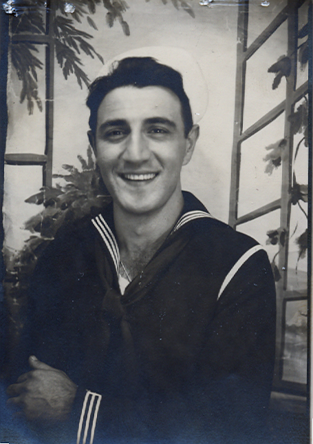 Erwin Hahn
A variety of campus researchers contributed to nuclear magnetic resonance, the field that predated MRI. The legacy began with chemistry Professor Herbert Gutowsky, who in 1948 became the first person to apply nuclear magnetic resonance to the
field of chemistry.
Erwin Hahn
A variety of campus researchers contributed to nuclear magnetic resonance, the field that predated MRI. The legacy began with chemistry Professor Herbert Gutowsky, who in 1948 became the first person to apply nuclear magnetic resonance to the
field of chemistry.
Erwin Hahn (M.S., '46; Ph.D., '49, physics) first detected spin echoes as a graduate student at Illinois. Spin echoes are a signal detected in a nuclear magnetic resonance spectrometer that can be used to extract more information from
a test sample than conventional nuclear magnetic resonance spectroscopy.
Interdisciplinary work leads to NMR breakthrough
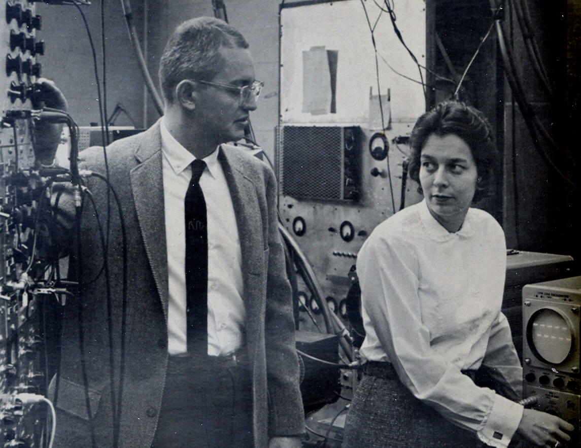 Physics Professor Charles Slichter pictured with then-graduate student Judy Franz (M.S., '61; Ph.D., ’65, physics).
Chemistry Professor Herbert Gutowsky and physics Professor Charles Slichter published "Coupling Among Nuclear Magnetic Dipoles in Molecules" in the journal Physical Review in 1951.
Physics Professor Charles Slichter pictured with then-graduate student Judy Franz (M.S., '61; Ph.D., ’65, physics).
Chemistry Professor Herbert Gutowsky and physics Professor Charles Slichter published "Coupling Among Nuclear Magnetic Dipoles in Molecules" in the journal Physical Review in 1951.
"The paper is one of two or three key papers in
the history of NMR spectroscopy that turned the technique from a curiosity into a method for determining the structure of molecules," said Martin Gruebele, the James R. Eiszner Endowed Chair in Chemistry.
Overhauser Effect developed on campus
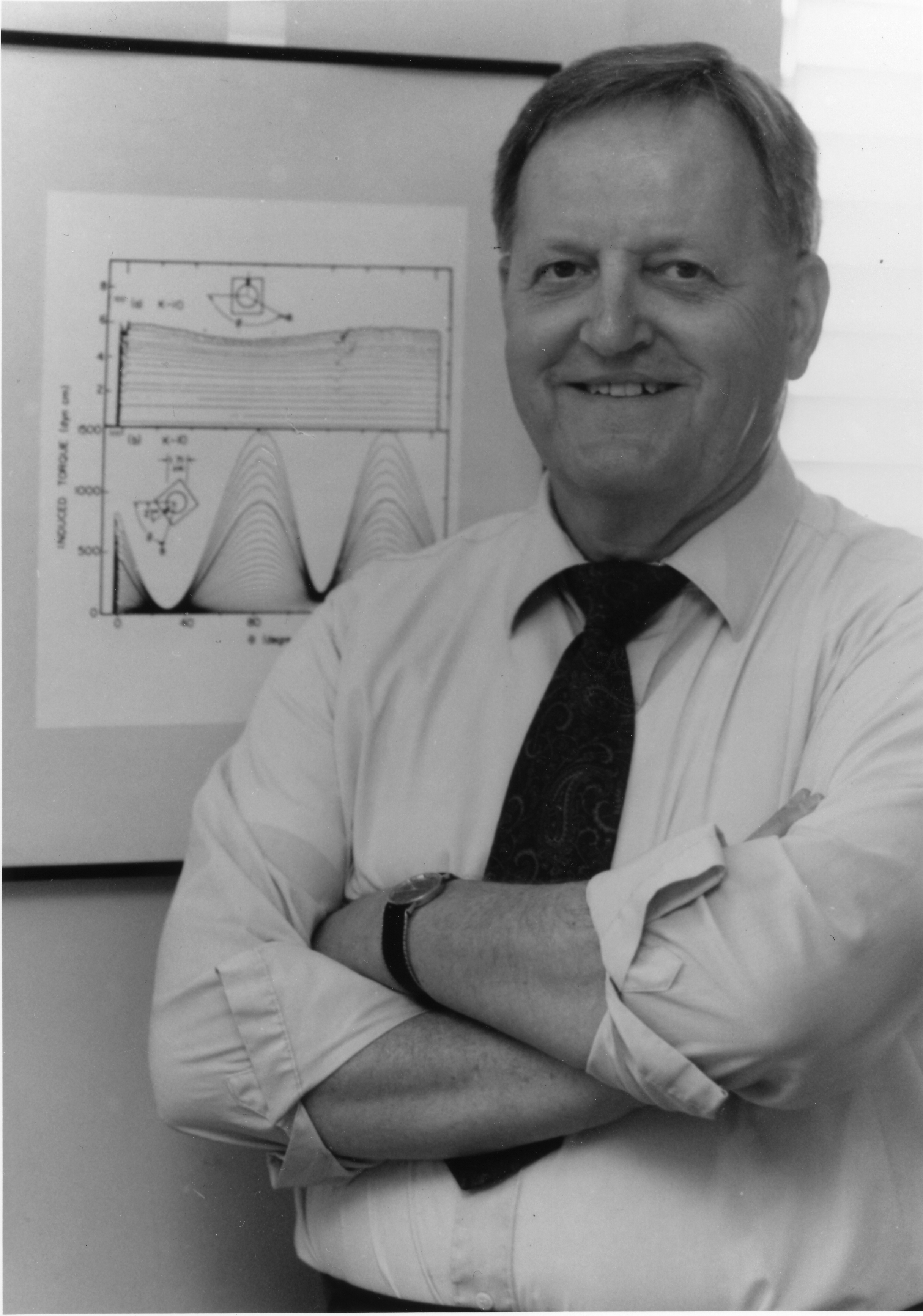 Albert Overhauser
In 1953, Illinois postdoctoral physicist Albert Overhauser developed the Overhauser Effect, a theory on the transfer of spin polarization. Slichter and his student, Tom Carver (Ph.D., ’54, physics), were the first to demonstrate the effect.
Albert Overhauser
In 1953, Illinois postdoctoral physicist Albert Overhauser developed the Overhauser Effect, a theory on the transfer of spin polarization. Slichter and his student, Tom Carver (Ph.D., ’54, physics), were the first to demonstrate the effect.
“The big shots in magnetic resonance did not believe Overhauser’s prediction, so it was a source of great excitement when we showed he was correct,” Slichter later said.
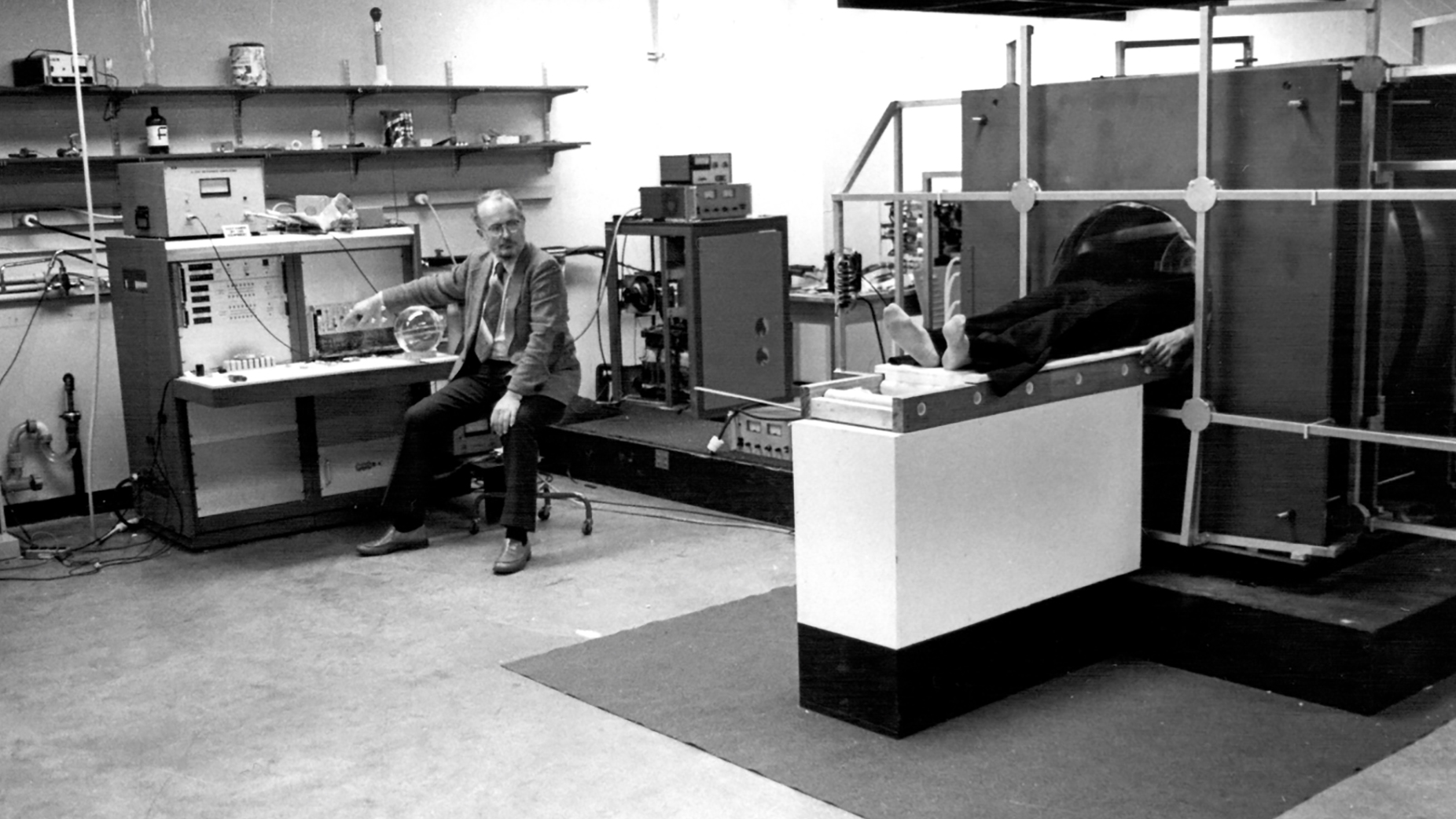
Lauterbur joins Illinois faculty
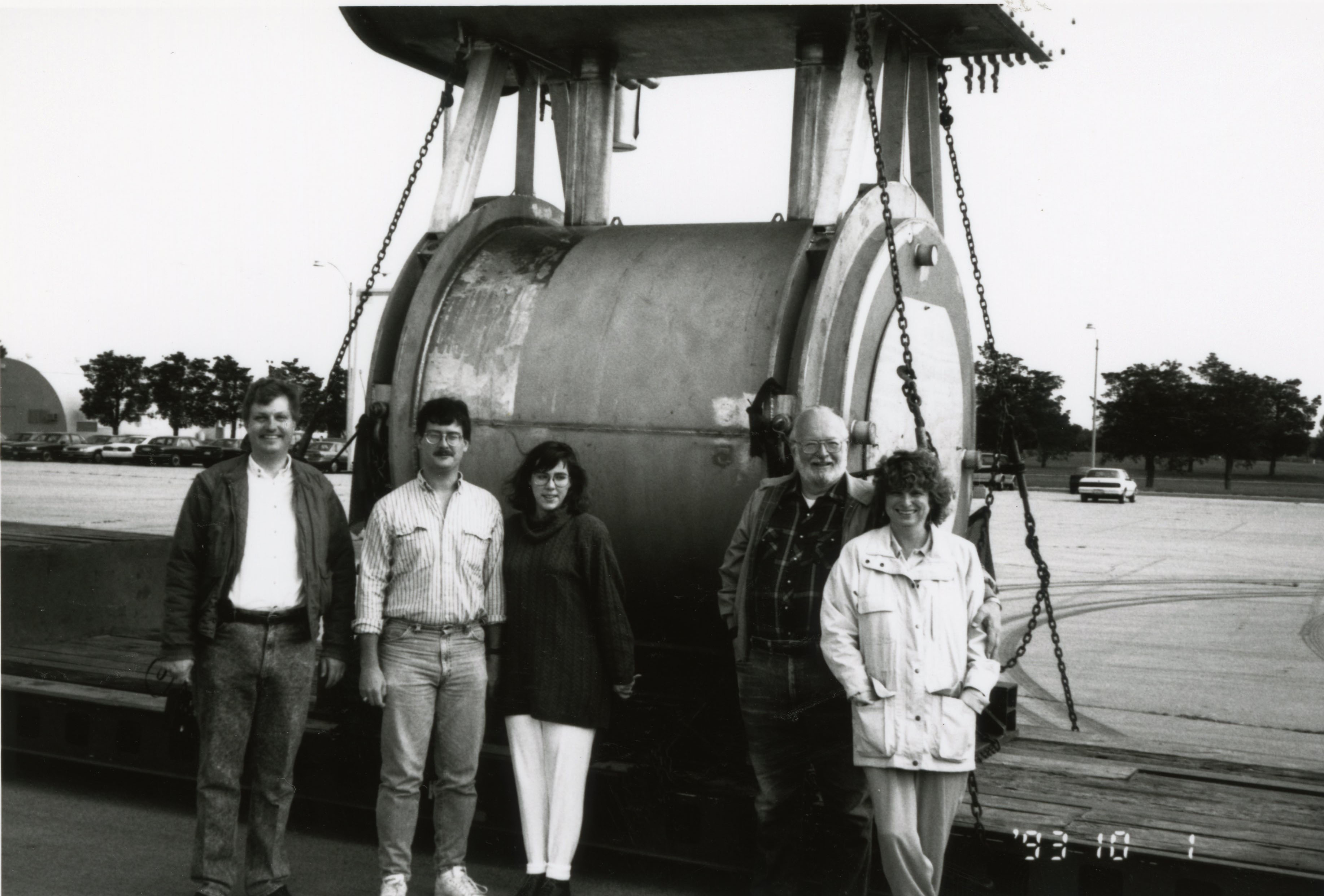 Paul Lauterbur, second from right, is pictured with members of the Biomedical Magnetic Resonance Laboratory with the MRI scanner funded by the National Science Foundation.In 1985, Paul Lauterbur joined
the University of Illinois faculty and served as the first director of the Biomedical Magnetic Resonance Laboratory.
Paul Lauterbur, second from right, is pictured with members of the Biomedical Magnetic Resonance Laboratory with the MRI scanner funded by the National Science Foundation.In 1985, Paul Lauterbur joined
the University of Illinois faculty and served as the first director of the Biomedical Magnetic Resonance Laboratory.
In 2003, he shared the Nobel Prize in Physiology or Medicine with Sir Peter Mansfield "for their discoveries
concerning magnetic resonance imaging." (Mansfield had previously performed magnetic resonance studies of doped metals as a postdoctoral researcher with physicist Charles Slichter on the Illinois campus.)
Lauterbur obtained the first magnetic
resonance image in 1971 at Stony Brook University.
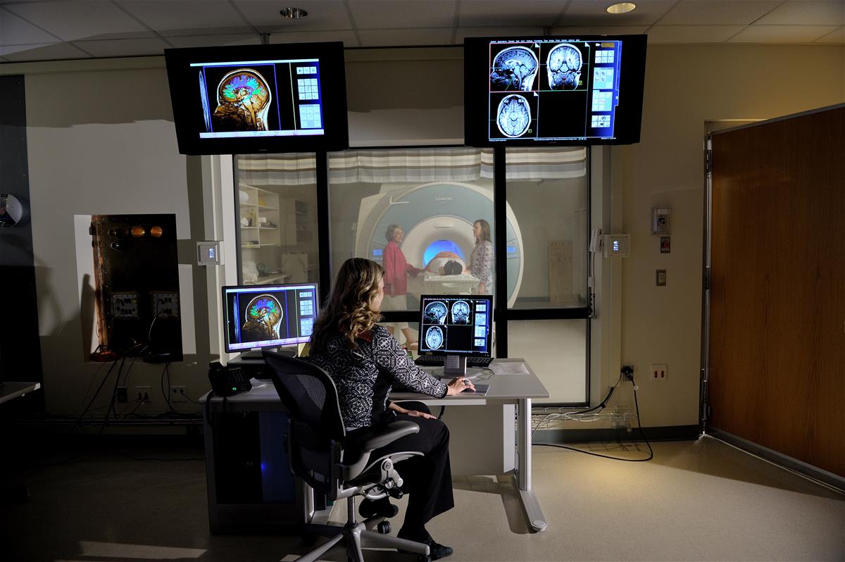
Biomedical Imaging Center drives research, innovation
 In 2002, the Beckman Institute established the Biomedical Imaging Center. This shared research facility helped drive innovation in MRI research, from developing new technologies to discovering ways to integrate other existing research tools. The
center provided opportunities for researchers in a variety of different fields to advance their research by taking advantage of new MRI techniques and technology.
In 2002, the Beckman Institute established the Biomedical Imaging Center. This shared research facility helped drive innovation in MRI research, from developing new technologies to discovering ways to integrate other existing research tools. The
center provided opportunities for researchers in a variety of different fields to advance their research by taking advantage of new MRI techniques and technology.
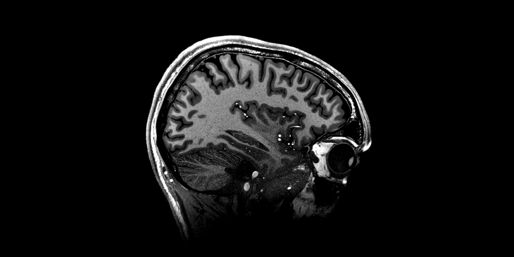
Current MRI research at the University of Illinois
Biomedical Imaging Center
 The Biomedical Imaging Center at the Beckman Institute is committed to the development of cutting edge techniques that combine magnetic resonance methods with other imaging
techniques, including optical imaging, eye-tracking, EEG, and more.
The Biomedical Imaging Center at the Beckman Institute is committed to the development of cutting edge techniques that combine magnetic resonance methods with other imaging
techniques, including optical imaging, eye-tracking, EEG, and more.
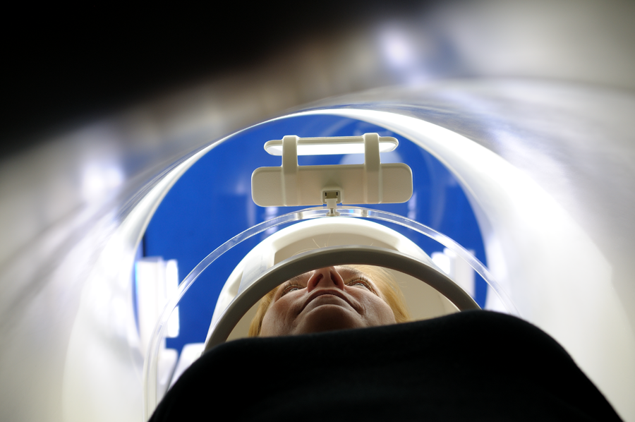
The center houses ultra-high field, molecular, and ultrasound imaging systems, as well as two state-of-the-art 3 Tesla MAGNETOM Prisma human whole-body MRI scanners. These systems include standard and custom head coils, knee coils, body
arrays, and even rat and piglet coils. For functional MRI studies, BIC has a full setup to enable investigators to perform high-quality studies.
Recent additions include a Bruker 9.4 Tesla preclinical animal MRI scanner and a shared Siemens MAGNETOM Terra 7 Tesla MRI housed at Carle Foundation Hospital.
9.4 Tesla animal MRI
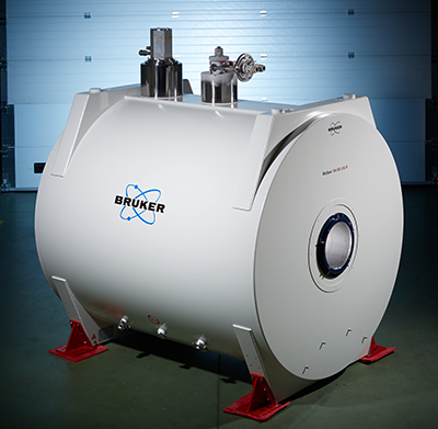 Eighteen University of Illinois colleges, departments, and schools, along with a generous gift from the Roy J. Carver Charitable Trust, funded the purchase
of a Bruker 9.4 Tesla preclinical animal MRI system for the Beckman Institute. The scanner further advances Illinois as an innovator in imaging technology.
Eighteen University of Illinois colleges, departments, and schools, along with a generous gift from the Roy J. Carver Charitable Trust, funded the purchase
of a Bruker 9.4 Tesla preclinical animal MRI system for the Beckman Institute. The scanner further advances Illinois as an innovator in imaging technology.
The new system delivers higher resolution imaging capabilities, enabling researchers
to analyze more intricate structures and processes in the brain, and advance their ability to recommend dietary and drug interventions to improve cognition, memory, behavior, and overall brain health.
Carle Illinois Advanced Imaging Center
 Carle Foundation and the University of Illinois Urbana-Champaign launched a first-of-its-kind partnership in 2019. One result: The purchase of a Siemens MAGNETOM Terra 7 Tesla MRI scanner.
Carle and the University of Illinois use its research capabilities with the scanner’s dual-mode functionality, which enables users to switch between clinical and research tasks, while storing clinical and research images on different databases.
“Working together, the two organizations conduct research at the frontiers of modern neuroscience — discovering new properties of the human brain,” said Tracey Wszalek, the director of the Beckman Biomedical Imaging Center and co-director
of the CIAIC.
Carle is one of the few clinical facilities nationwide to offer this advanced technology to patients. The scanner is located at Carle and represents the first such scanner to be placed in a community health setting.
The scanner provides the highest magnetic field imaging strength commercially available in the U.S. approved by the Food and Drug Administration for brain and knee scans.
Together, Carle and UIUC have created the C-U Population Study, a comprehensive look at brain and cognitive health through 7 Tesla imaging.
Harnessing new technology in biomedical imaging
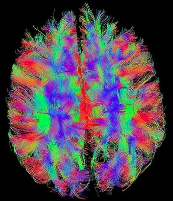 Magnetic resonance imaging has been an important scientific tool for decades. In recent years, newer biological imaging tools have become increasingly useful in a wide range of disciplines.
These imaging tools provide never-before-seen
views of the biological world — both for scientific research and for medical purposes, such as diagnosis and therapy.
Beckman Institute researchers are at the forefront of this imaging revolution.
Beckman researchers
both develop and use functional MRI to link neural physiology to cognitive function by measuring blood flow in activated areas of the brain.
The researchers are advancing ultrasound techniques, and using them for things like visualizing the distribution of blood vessels and measuring oxygen levels in tumors and understanding ultrasonic communications with implanted medical devices through tissue.
Other institute researchers are developing their own distinct technologies and processes. For example, those working with optical coherence tomography are studying how to use light waves to instantly and noninvasively image tissue. And others work with spatial light interference microscopy, which can measure dynamics in live cells.
Many multimodal techniques — such as simultaneously combining MRI with optical imaging and the electrical signals generated by the brain — were pioneered at Illinois.
MRI news
- Beckman researchers map prefrontal cortex in individuals, reveal fine-scale structure with precision fMRI
- Team comprehensively maps brain with single scan
- Beckman researchers to model brain, better understand traumatic brain injuries
- Beckman announces 2024 research seed grant awardees
- Brad Sutton named an ISMRM Fellow
- Beckman’s Owl Patrol takes flight
- Webb makes MRI work for research, clinics, and communities worldwide
- Seven things to know about the 7 Tesla MRI scanner
- MRI turns 50: Expert Brad Sutton explains its history and role in understanding the aging brain
- Making the most of MRI: Beckman welcomes visiting scholar, MRI expert Luisa Ciobanu
Beckman Institute for Advanced Science and Technology