Article
“Small-molecule Probe Development for Photoacoustic Imaging of Prostate Cancer Hypoxia”
Hailey Knox, a Ph.D. student in chemistry and a Beckman Institute Graduate Fellow
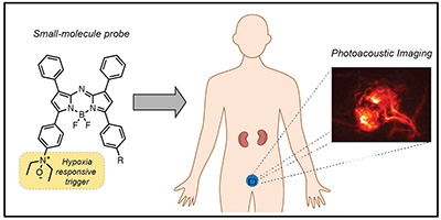
Prostate cancer is the most common cancer diagnosis in men, with more than 100,000 new cases appearing each year and over 3 million survivors in the United States. While the five-year survival rate for prostate cancer patients diagnosed at an early (non-metastatic) stage is nearly 100 percent, those who are diagnosed after the cancer has spread face a mere 29 percent survival rate, indicating the critical importance of early detection. Current methods for prostate cancer screening are unreliable and often lead to false positives and unnecessary treatments. Additionally, prostate cancer staging is performed through the use of invasive biopsy procedures that require removal of prostate tissue and are unable to report on the entire volume. Because accurate staging of prostate cancer leads to effective treatments and reduced mortality, it is essential to seek new diagnostic approaches that can reliably report on prostate cancer stage. Hypoxia is a key characteristic of prostate tumors and is correlated with clinical stage. Our work seeks to utilize prostate tumor hypoxia as a biomarker for diagnostic and prognostic applications. Specifically, we aim to develop small-molecule probes that can be used for photoacoustic (PA) imaging of prostate cancer hypoxia. PA imaging is a powerful imaging modality that combines the high resolution of optical imaging with the depth penetration of ultrasound and can be used in conjunction with optically-absorbing dyes to report on specific targets or microenvironments. To accomplish this, we have developed an N-oxide-based chemical trigger that can alter the PA output of a small-molecule dye in response to tissue hypoxia. The utility of this molecular design has been demonstrated for hypoxia imaging in cell culture and in murine models of cancer and acute ischemia. We have also employed chemical tuning to optimize the photophysical properties and PA signal of our small-molecule probes, leading to new designs with brighter signal and greater sensitivity. Our current work is aimed at efficiently targeting probes to prostate cancer tissue to increase sensitivity for hypoxia detection. By employing high-resolution imaging with molecular probes, we hope to enable acquisition of key prognostic information in a completely non-invasive manner.
“Neuropeptides: The Keys to Unlock Chronic Pain Mechanisms"
Krishna D.B. Anapindi, a Ph.D. student in applied statistics and a member of Beckman's Neurotechnology for Memory and Cognition Group
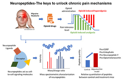
Migraine is one of the most debilitating neurological disorders which affects over 40 million people annually in the United States alone. Patients suffering from migraine are often prescribed opioids as a frontline medication to alleviate their pain. However, persistent use of opioids leads to a paradoxical situation known as opioid-induced hyperalgesia (OIH), where the subject administered with opioids becomes more sensitive to a pain stimulus. Moreover, opioid-based therapies are highly addictive, and their abuse has led to a devastating public health crisis. Despite having such an enormous impact on the health status of the population, precise molecular mechanisms driving migraine and OIH are still not well understood. To gain insights into molecular underpinnings of these multifactorial pain conditions, specifically neuropeptide dysregulation in migraine and OIH rodent models, we combine physiology, behavioral pharmacology, and omics technologies in the study unusual in its scale and ambition. Leveraging the power of label-free mass spectrometry in unbiased and untargeted peptide characterization, we were able to correlate the change in levels of several neuropeptides including CGRP[83-119] (Calcitonin gene-related peptide), NPY[29-64] (Pro-Neuropeptide Y), TKN[101-107] (Pro-Tachykinin) and PENKB[166-170] (Pro-EnkephalinB/Pro-Dynorphin) in these two conditions. Furthermore, we found the composite peptide complement of the prohormone PACAP to be strongly correlated with both migraine and OIH. In Addition, we performed a further pharmacological analysis and confirmed the involvement of PACAP in bridging these two pain disorders. We are hopeful that the peptide and protein candidates identified in this study will help elucidate the mechanistic processes underlying these conditions and eventually help develop a reliable and robust cure.
“Compressed Ultrafast Photography and Microscopy: Redefining the Limit of Passive Ultrafast Imaging"
Yayao Ma, an M.S. student in electrical and computer engineering and a Beckman Institute Graduate Fellow
Beckman Institute for Advanced Science and Technology