Article
“Hydrophobicity-driven Unfolding of Trp-cage Encapsulated between Graphene Sheets”
Zhikun Cai, nuclear, plasma, and radiological engineering (NPRE); Beckman Institute Graduate Fellow; Computational Molecular Science (CMS) Group
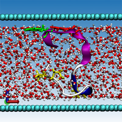
Understanding the interaction between proteins and graphene not only helps elucidate the behaviors of proteins in confined spaces but is also imperative to the development of a plethora of graphene-based biotechnologies. To study the overall geometrical-thermal effects on proteins, we performed molecular dynamics simulations of hydrated Trp-cage miniprotein sandwiched between two graphene sheets and in the bulk environment at the temperatures below and above its unfolding temperature. We observed that at both temperatures the confined protein became adsorbed to the graphene surfaces and exhibited unfolded structures. Residue-specific analyses clearly showed the preference for the graphene to interact with the hydrophobic regions of Trp-cage. These results suggested that the conformation space accessible to the protein results from the interplay between the geometrical restraints and the thermodynamic driving forces. While confinement usually tends to restrict the conformation of proteins by volume exclusion, it may also induce the unfolding of proteins by hydrophobic interactions.
“High Definition and Fast Infrared Imaging of Breast Tissue: A Paradigm Shift in Cancer Diagnosis and Survival Predictions”
Shachi Mittal, bioengineering (BioE), Beckman Institute Graduate Fellow, Bioimaging Science and Technology Group
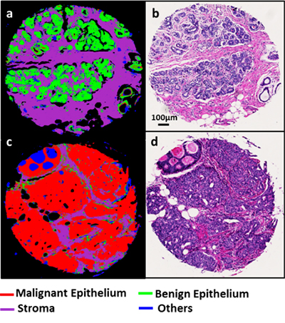
Breast cancer survival rates and prognosis is highly dependent on the stage of tumor, making early detection vital for improving patient outcome. The existing methods being used for clinical predictions are either lacking in the morphological or biochemical detail. Infrared (IR) imaging combines these two information levels to provide spatially resolved and quantitative information of biological samples. Currently, breast cancer diagnosis is limited by a large number of false positives and low throughput research. Fourier Transform Infrared (FT-IR) Spectroscopy, on the other hand, can be used to harness the encoded biochemical information in patient samples in an automated manner. The long term goal is to develop clinically translatable automated protocols by integrating concepts from different disciplines toward a post screening and a diagnostic tool for breast cancer detection. Our preliminary data analysis of 100 tissue samples and 47 patients suggests that high-definition (HD) IR imaging can distinguish between diseased states and different stromal configurations with an area under the receiver operating characteristic curve of more than 0.95.The rationale that underlines the proposed research is that the IR spectra from diseased and normal physiology provides distinct chemical information of a tissue sample which can be used to conduct digital pathology.
“Photoacoustic Microscopy Using Photonic Integrated Circuit Detectors”
Jorge Tordera Mora, electrical and computer engineering (ECE), Bioimaging Science and Technology Group
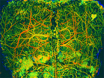
Photoacoustic microscopy (PAM) is a powerful technique able to break the optical diffusion limits by combining optical excitation and acoustic detection. Since the acoustic scattering coefficient is three orders of magnitude less than optical scattering through tissue, PAM can achieve high-resolution images at greater depths than common microscopy techniques. Optical excitation is produced by nanosecond laser pulses focused onto biological tissues where light is absorbed, leading into a rise of temperature, thermal expansion, and acoustic emission that can be detected by high-frequency ultrasound devices. Then, by repeating this process in the form of a raster scan, flight time of the ultrasound signal provides depth information (3D image). Multiple acoustic detection devices from conventional transducer to Photonic Integrated Circuit Detectors (PICs)—such as microspheres or micro-ring resonator—have been developed in order to improve constraints in size or opacity of transducers providing highly sensitive detection, broader ultrasonic detection bandwidth and large detection angle. This talk will highlight the unique advantages of a PAM setup and its state-of-the-art detection devices.
Beckman Institute for Advanced Science and Technology