Article
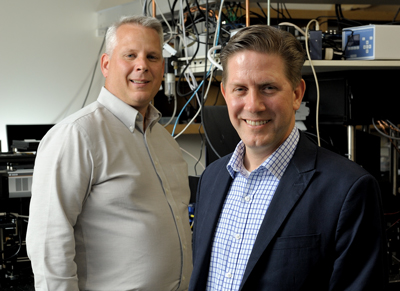 Co-directors for the GSK Center for Optical Molecular Imaging are Zane Arp, left, U.S. lead for imaging technologies at GSK, and Stephen Boppart, a professor of electrical and computer engineering and of bioengineering, and co-chair of the Integrated
Imaging theme.
Co-directors for the GSK Center for Optical Molecular Imaging are Zane Arp, left, U.S. lead for imaging technologies at GSK, and Stephen Boppart, a professor of electrical and computer engineering and of bioengineering, and co-chair of the Integrated
Imaging theme.
The research represents a unique Beckman collaboration—a partnership between Beckman’s Biophotonics Imaging Laboratory and the pharmaceutical company GSK, which created the GSK Center for Optical Molecular Imaging in 2016.
Perfect match
GSK’s global search for the right partner lead them to the Biophotonics Imaging Lab at the Beckman Institute and the lab’s director, Stephen Boppart.
“Steve proposed a structure where we have GSK working side-by-side in the lab with postdocs and grad students working on the same project,” said Zane Arp, U.S. lead for imaging technologies at GSK and co-director of the center. “They’re advancing the technology. We’re advancing the applications. And the marriage of those two makes the sum greater than either part.”
“Our advanced optical imaging techniques also enable new molecular, metabolic, structural, and functional imaging of cells and tissues, even in human subjects,” said Boppart, who is co-chair of the Integrated Imaging theme and co-director of the GSK Center. “All of these aspects were directly relevant to what GSK was looking for in order to advance their drug discovery and development. There is a clear need to image drug distribution and efficacy at the molecular-cellular level in living tissues, and our techniques and technologies were a perfect fit.”
Improving efficacy
The techniques involve laser light that penetrates the skin about 200-500 microns. (The width of a single human hair ranges from about 50-100 microns.) The light can actually penetrate completely through the skin—up to 10 centimeters—but it only provides valuable high-resolution imaging and feedback at about 200 microns in depth, according to Arp.
“Traditional imaging modalities such as MRI, x-ray, and CT typically measure morphological (or structural) changes,” Arp said. “We’re looking more for the small changes, the chemical-level changes that occur before you ever see that morphological change.”
Detecting how medications work at the molecular-cellular level can save time and money.
The new optical imaging techniques allow researchers to see what targets the medication is hitting in a variety of different systems including cells, animals, and humans. This allows vertical translation of methods from these systems into clinical work, such as dermatology. In dermatology, the techniques can be used to determine how long the drug stays on the skin, the dosing regimen, and drug-target interaction. Skin is the body’s natural protective layer so figuring out how much medication—if any—is penetrating the skin can be problematic.
“Most dermal drugs fail because they don’t manage to get through the stratum corneum (the first layer of skin),” said Aneesh Alex, who is a GSK employee and a postdoctoral fellow in the center.
“As a drug development company, one of the critical aspects of our dermatologic drugs is how we defeat the barrier so that we’re getting our medicine where it needs to go in the patient,” Arp said.
“If you get too much penetration too quickly, you’ll just wash it out into your bloodstream,” he explained. “If you get too little penetration then you don’t see any effect. We also have to prove that the drugs hit the primary target the hardest. That’s where we get the most efficacy. It’s an art actually.”
Another benefit of the new platform of technologies is that they allow researchers to observe changes at the cellular level without tagging the medication applied with a dye, which can impact the test results.
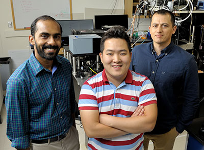 Three GSK Postdoctoral Fellows—from left, Aneesh Alex, Jang Hyuk Lee, and Jose Rico-Jimenez—assist with research at the GSK Center for Molecular Optical Imaging based at the Beckman Institute.
Getting results
The center has three to four projects in progress, and researchers have completed nearly half-a-dozen studies with several in publication.
The noninvasive techniques also allow the center to do its dermatological research on healthy human subjects as well as animal models.
“The other beauty of this is that we can do what we call an optical biopsy because it eliminates the need for a true biopsy, where you need to extract a tissue sample,” Arp said.
The center also benefits from additional services and facilities at the institute.
“Another advantage is the facilities at Beckman, including the Microscopy Suite and other imaging facilities,” Alex said. “In the future, we are also planning to go beyond the optical capabilities so we want to make use of the MRI and PET capabilities that we have here. And I think that is something unique about Beckman. We have all the resources here under one roof. I think that is one of the main reasons GSK wanted to come here. Not a lot of places have these kinds of resources in one location.”
And even though it’s early, the center is seeing results and impacting decisions at GSK that will improve patient
health care.
“Being able to see our drugs have an action at the earliest possible time point improves safety, improves the efficacy, and improves our knowledge base,” Arp said.
Alex also touted the benefits to the students involved in the research.
“I think by being able to be a postdoc here, I am getting the best of both worlds,” he said. “Because I see the industrial aspect and also the academic aspect, it’s really a unique development opportunity."
“We are confident that we are on the right track, and that we have a very unique academic-industry partnership that is demonstrating early success,” Boppart said. “I believe it is important to continue this work because of the significant impact it can have, not only on drug development, but also ultimately on patient health and health care.”
“We’re very pleased with the progress being made,” Arp said. “We have had senior managers come out here who are extremely pleased with the type of work we’re doing and the fact that we’re getting value out of cutting-edge
capabilities five to
10 years before the techniques are commercially available is a big deal.”
The center also is helping to drive the future of technology. For one, reducing the size of the equipment in order to help diagnose and treat individual patients is a possible long-range goal.
“We expect that we will start seeing biomarkers that are specific to individual patients or patient populations,” Arp said. “But I would say that using these technologies to differentiate patient populations for personalized medicine is a larger goal for us at some point. That’s something we see out in the future.”
Beckman Institute for Advanced Science and Technology