Article
“The Cardiotoxic Implications of Modulating CYP2J2 Arachidonic Acid Metabolism”
William Randall Arnold, graduate research assistant, 3D Micro- and Nanosystems Group
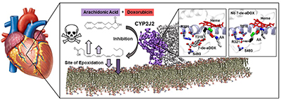
Polyunsaturated fatty acids like arachidonic acid (AA) are gaining scientific interest insofar as they are converted in the body by cytochrome P450 2J2 (CYP2J2) to cardioprotective mediators. These mediators, termed EETs, regulate heart rhythm; vasodilation, preventing ischemic injury and hypertension; prevent cardiac cell death; and overall prevent cardiac diseases. CYP2J2 also metabolizes several cardiotoxic drugs, and it has been hypothesized that these drugs are cardiotoxic because they disrupt CYP2J2’s production of EETs. We have shown, for the first time, that a cardiotoxic drug, doxorubicin (DOX) directly modulates CYP2J2-mediated AA metabolism. DOX is known to produce reactive oxygen species (ROS) to induce toxicity, however, the mechanisms governing DOX’s particular cardiotoxicity are unknown. We determine experimentally and through molecular dynamics simulations that DOX inhibits CYP2J2’s AA metabolism. Furthermore, we determine that a metabolite of DOX, 7-deoxydoxorubicin aglycone (7-de-aDOX), changes the site of metabolism of AA. Together, these data suggest that DOX produces an initial assault to cardiac cells partly through the production of ROS. DOX then reduces the ability for cardiac cells to combat this assault by inhibiting the production of EETs. Finally, as DOX is converted to 7-de-aDOX, 7-de-aDOX changes the site of AA metabolism, which may further cardiotoxicity.
“Impact of Zika Virus on Adult Human Brain Structure and Functional Organization: a Collaboration with the Dominican Republic
Richard O. Bido-Medina, M.D./PhD Candidate, Intelligence, Learning, and Plasticity Group
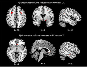
Since its outbreak in the Americas in 2015, Zika virus (ZIKV) has become a major global health threat due to its potential to affect the nervous system. Although most adult cases show no or mild symptoms, some patients exhibit severe neurological complications. While these complications often largely present as Guillain-Barré Syndrome (GBS)-like manifestation confined to the peripheral nervous system (PNS), they include symptoms of the central nervous system (CNS) in a subset of severely affected patients. Yet, reports on the adult CNS have remained confined to few single cases of structural brain changes, and no functional neuroimaging studies have investigated how ZIKV affects adult human brain function. In this first case-control neuroimaging study, we investigated nine rare adult patients with ZIKV-related neurological complications of the CNS during the subacute phase (3 females, Age=35±4.46, range 30-45 years, 5±0.5 months since onset of symptoms). Patients showed both structural and functional brain changes compared to nine healthy age- and sex-matched controls. Results show that ZIKV can affect adult human brain structure and functional organization, comprising both motor-related regions likely secondary to prolonged PNS weakness, as well as nonsomatomotor regions indicative of PNS-independent alternations. In the context of the observed unusual non-ascending presentation of GBS-like symptoms in ZIKV patients, our clinical and neuroimaging observations could indicate that GBS due to ZIKV is an atypical variant that, in severe cases, may extend to the CNS. Our observations highlight an urgent need for future studies to understand ZIKV-related neuroinflammatory mechanisms in adults, potentially comprising direct CNS infections or autoimmune processes influencing the CNS.
“LiMn2O4 Surface Chemistry Evolution During Cycling Revealed by in situ Auger Electron Spectroscopy and X-ray Photoelectron Spectroscopy”
Jenny Beach, graduate research assistant, 3D Micro- and Nanosystems Group
This work utilizes in situ electrochemical and analytical characterization during cycling of LiMn2O4 (LMO) equilibrated at different potentials in an ultrahigh vacuum (UHV) environment. The LMO reacts with organic molecules in the vacuum to form a high surface concentration of Li2CO3 (≈50% C) during initial charging to 4.05 V. Charging to higher potentials reduces the overall Li2CO3 concentration (≈15% C). Discharging to 3.0 V increases the Li2CO3 concentration (≈30% C) and overdischarging to 0.1 V again reduces its concentration (≈15% C). This behavior is reproducible over five cycles. The model geometry utilized suggests that oxygen from LMO can participate in redox of carbon, where LMO contributes oxygen to form the carbonate in the solid-electrolyte interphase (SEI). Similar results were obtained from samples cycledex situ, suggesting that the model in situ geometry provides reasonably representative information about surface chemistry evolution. Carbon redox at LMO and the inherent voltage instability of the Li2CO3 likely contributes significantly to its capacity fade.
Beckman Institute for Advanced Science and Technology