Article
With a keen eye and the correct tools, a science experiment can become anything.
The seven winners of this year's Beckman Institute Research Image Contest used smartphones, microscopes and their own artistic perspectives to transform research projects into pearl necklaces, outer-space expeditions, deadly wildlife encounters and more.
The contest is open to researchers affiliated with the Beckman Institute for Advanced Science and Technology as well as non-Beckman researchers on campus who use the institute's core facilities. This year's winners include undergraduate and graduate students, postdocs, staff and faculty members.
"I'm very pleased that so many of our researchers found so much beauty in their work and wanted to share it," said Catherine Murphy, the former Beckman Institute interim director at the time of this contest. "We had a tough time picking the winners. Congratulations to all!"
A team of Beckman administrators, staff and faculty members blind-judged the entries on their visual and research appeal. The winning images will be displayed for one year in the Beckman Institute East Atrium and one year in the Beckman Director's Conference Room before finding permanent homes across the institute and in the community.
This year's winners are:
Undergraduate student category
Visualizing the unseen
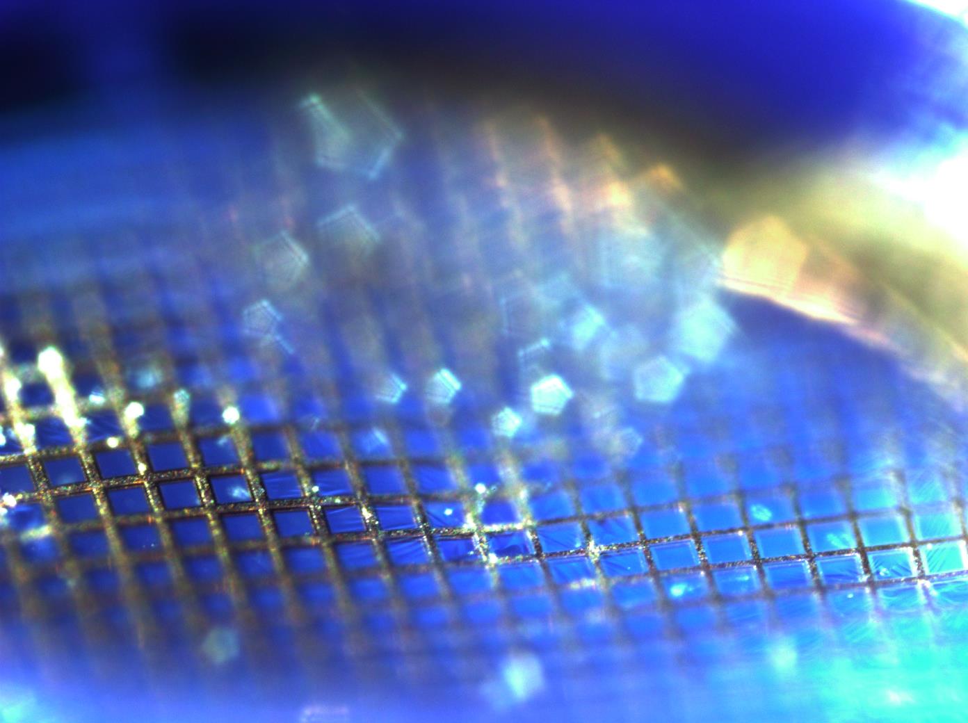 Visualizing the unseen. Credit: Diana Pham.
Metal nanoparticles magnified to 100 times their original size appear as floating pentagons due to the angled, intensely focused light on the pointed upper edges of the metal grid lines. Because the nanoparticles are too small for optical microscopes, which magnify objects with visible light, researchers turn to transmission electron microscopes, which magnify objects with fast-moving electrons.
Diana Pham is an undergraduate student in the Department of Mechanical Science and Engineering. At Beckman, she works with chemistry professor and former Beckman Institute Interim Director Cathy Murphy; 2023 Beckman-Brown Interdisciplinary Postdoctoral Fellow Kelly Powderly; and Joe Maduzia, a principal research engineer at the Micro-Nano-Mechanical Systems Laboratory. This image was captured with a 100 times magnification microscope in the Micro-Nano-Mechanical Systems Cleanroom Laboratory within the Department of Mechanical Science and Engineering.
“The research represented by this image will help researchers interested in analyzing gold, silver and copper nanoparticles. This could help people with climate change applications to further understand how nanoparticles are distributed and modified in complex biological systems,” Pham said.

Graduate student category
Into the polymer nanoverse
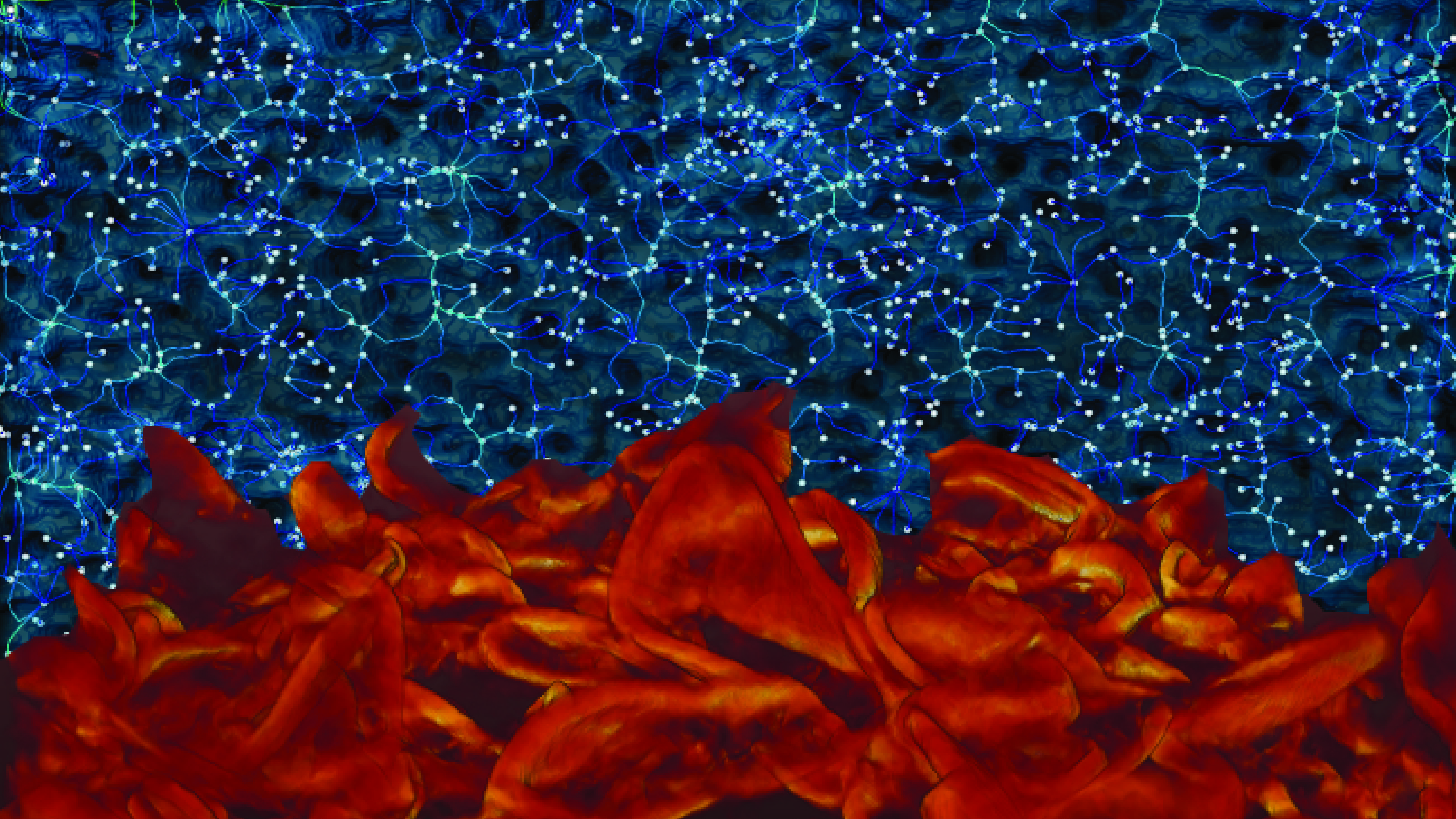 Into the polymer nanoverse. Credit: Falon Kalutantirige.
Polymer filtration films are used in water purification to separate impurities from drinking water. My research investigates how the films’ irregular architecture (i.e., structure and thickness) affects how well they work. Here, 3D renderings of polymer films resemble land surfaces and star constellations. I used a 3D thickness map to illustrate a hot, mountainous planet and a 3D network map to simulate a dark, starry universe.
Falon Kalutantirige is a graduate student in the Department of Chemistry working with Qian Chen, a Beckman researcher and professor of materials science and engineering. This image was reconstructed using software in Beckman’s Visualization Laboratory.
“Our work will help develop more efficient filtration films, eventually providing access to cleaner drinking water," Kalutantirige said.
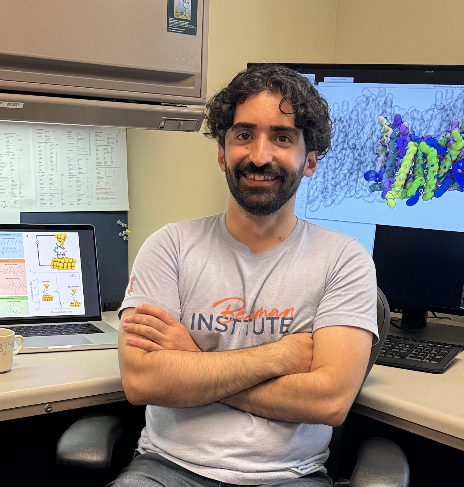
Do peptides dream of electric sheep?
This image received the school choice award from students at Champaign’s Edison Middle School and will be displayed in the school building during the 2024-2025 academic year.
 Do peptides dream of electric sheep? Credit: Moeen Meigooni.
Proteins inside our cells keep our bodies balanced and healthy. For example, they help create an energy source called ATP, which enables us to do things like move our muscles. This computer model shows an energizing circuit inside a protein, where electrons (the streak of electricity) cross a peptide power line (the tan, white, red and blue structure in the middle) between two gold electrodes.
Moeen Meigooni is a graduate student in the Center for Biophysics and Quantitative Biology. At Beckman, he works with Emad Tajkhorshid in the Theoretical and Computational Biophysics Group and collaborates with Charles Schroeder, a professor of materials science and engineering. This image was captured using Blender and VMD, a molecular visualization software developed at Beckman. Additional credit: Rajarshi Samajdar.
"This research enhances our understanding of how energy flows through proteins, offering deeper insights into fundamental cellular processes such as respiration and photosynthesis and accelerating the development of bioinspired technologies for energy and nanotechnology applications,” Meigooni said.
Postdoc and staff category
The deadly bite
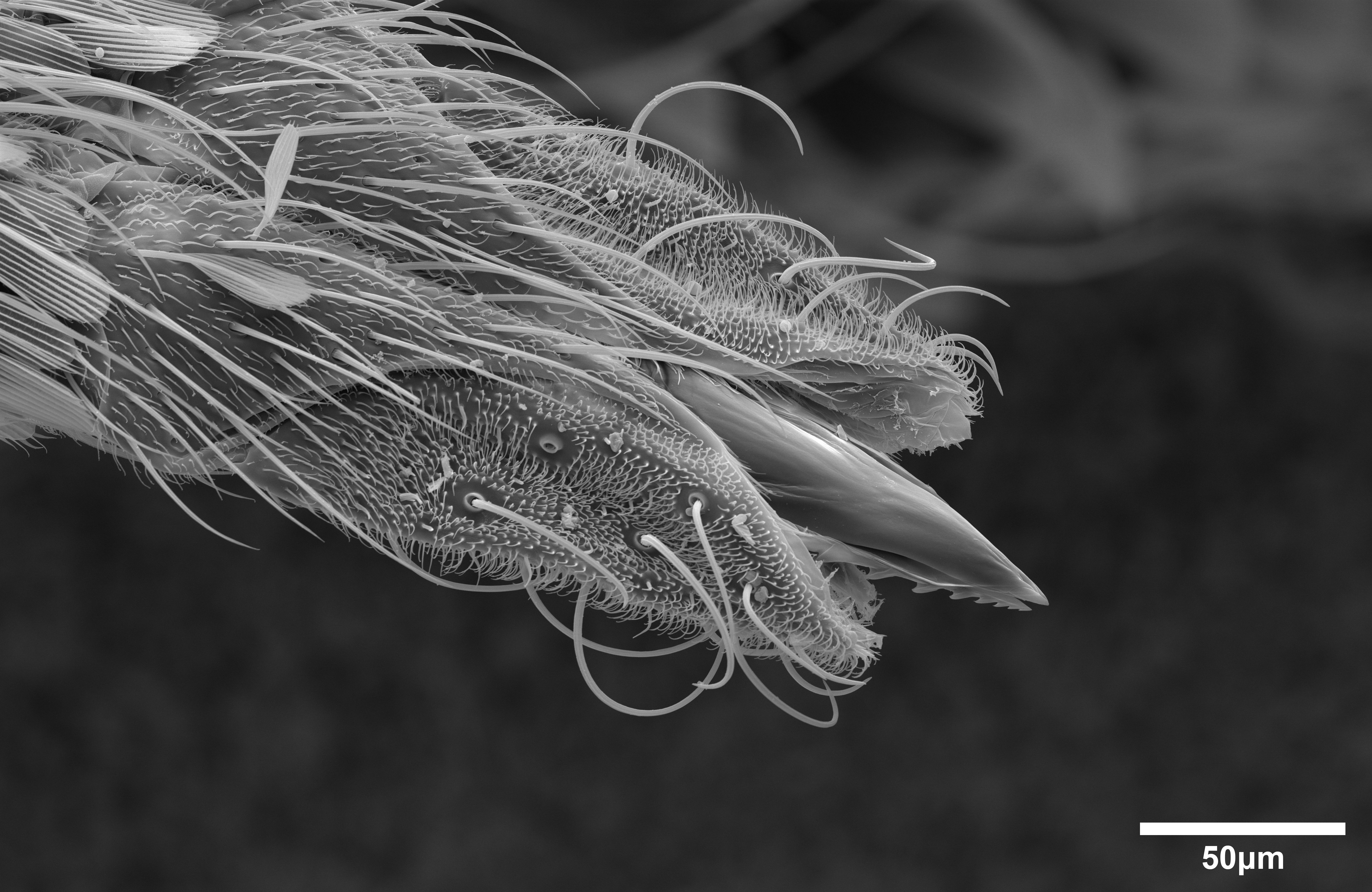 The deadly bite. Credit: T. Josek.
Known for sucking blood and spreading disease, mosquitoes are one of the world's deadliest animals. Here you can see a mosquito’s serrated mouthparts, or maxillae. Typically, the maxillae are hidden in a protective sheath and are only exposed right before a mosquito bites its host, making this a rare and terrifying view.
T. Josek joined the Beckman Institute in 2022. They are a microscopist in Beckman’s Microscopy Suite, one of the institute’s core imaging facilities. This image was captured with an environmental scanning electron microscope.
“Mosquitoes are a huge health concern, especially for humans. This image helps people see the mechanism mosquitos use to pierce our skin, which is typically thought to be more like a straw as opposed to the serrated, needle-like mouthparts seen here,” Josek said.

Golden necklace
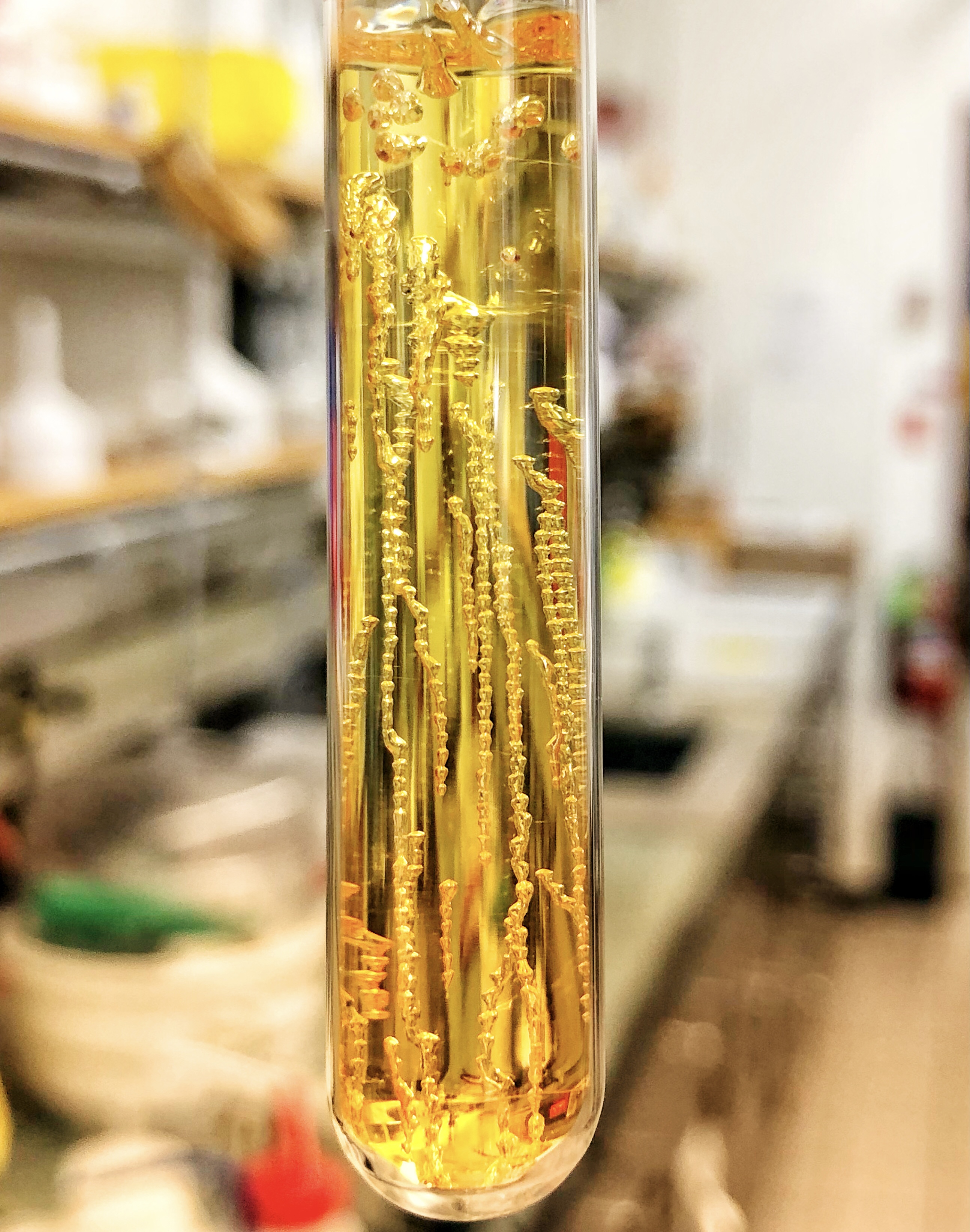 Golden necklace. Credit: Xiaolin Liu.
Frontal ring-opening metathesis polymerization, or FROMP, is an energy-saving chemical reaction that uses a small amount of heat to harden a soft material into a plastic. To create this golden resin, we applied heat to a colorless chemical called dicyclopentadiene and added the ruthenium Grubbs catalyst, which speeds up the reaction and gives the resin its golden color. The strings of bubbles, which look like jewelry, formed because of trapped air and water pockets vaporized by the heat.
Xiaolin Liu is a postdoctoral researcher working with Jeff Moore, a research professor in chemistry. The image was captured with Liu’s iPhone X.
“Our research aims to produce strong, durable polymer materials using as little energy as possible, reducing the energy footprint of plastic production and propelling engineering toward a more sustainable future,” Liu said.
Faculty category
Seeing the nanoworld
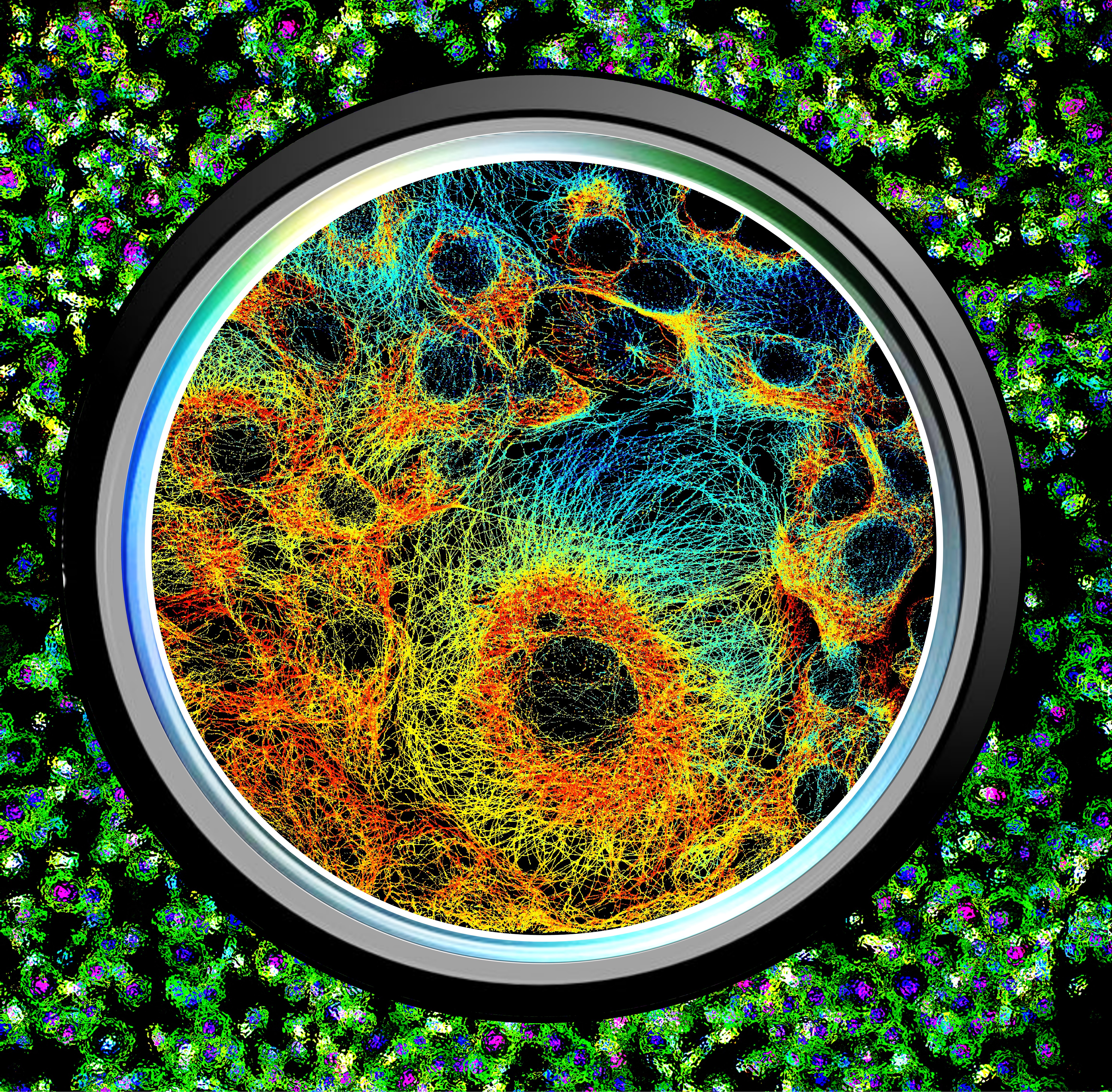 Seeing the nanoworld. Credit: Hongqiang Ma.
This 3D super-resolution image showcases the microtubules of human cells, offering a spatial resolution of 20 nanometers. The image encompasses around 200 million individual molecules, captured by our ultra-high throughput super-resolution imaging system in a mere 200 seconds.
Hongqiang Ma is a research professor in bioengineering. At Beckman, he collaborates with Yang Liu in the Biomedical Optical Imaging Laboratory. This image was obtained using laboratory-built, high-throughput super-resolution microscopy.
“Our research benefits medical professionals by enabling high-precision early diagnosis, treatment and understanding of diseases at the molecular level, which enhances patient outcomes and quality of life. It also assists scientists across disciplines in visualizing biological structures and molecular interactions with unprecedented detail, leading to breakthrough discoveries and advancements in knowledge,” Ma said.

Cellular landscape of the mammal brain
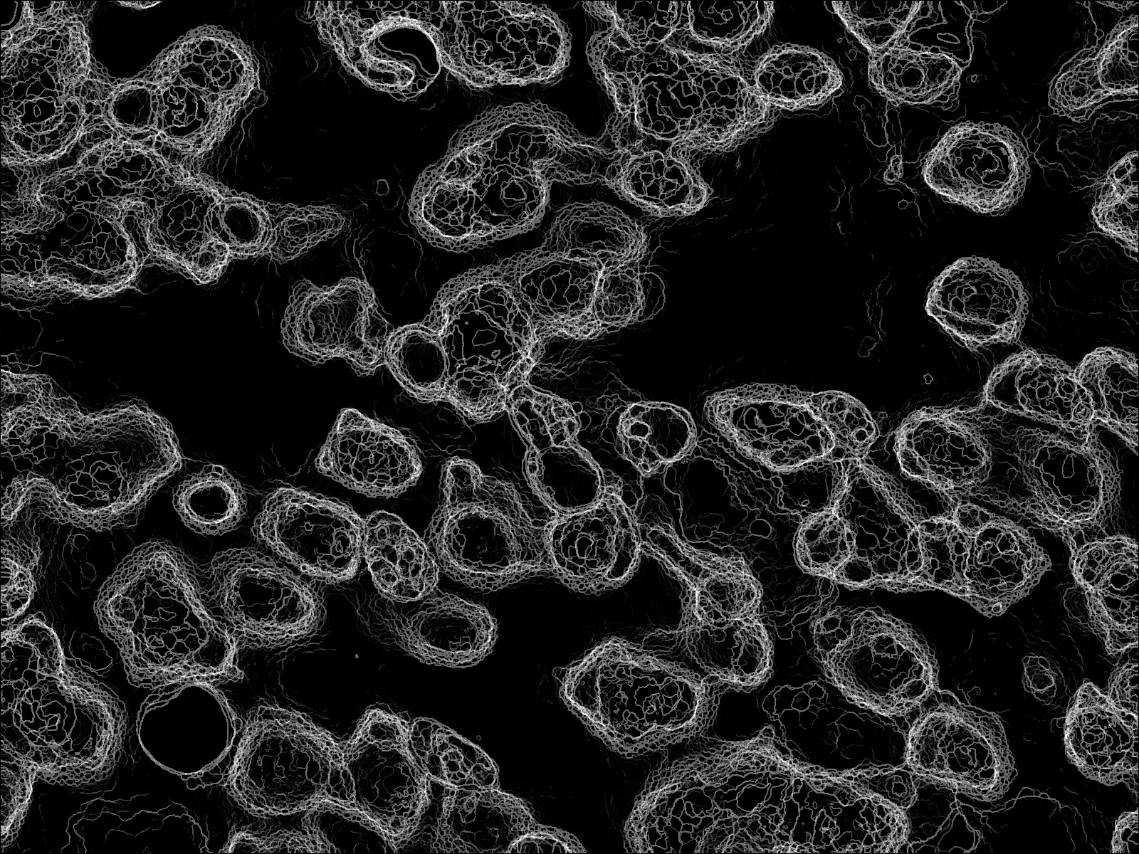 Cellular landscape of the mammal brain. Credit: Dan Miller.
This image depicts the topographic landscape of brain cells. The cells’ edges appear in white against a black background. This image forms the basis for our research, which aims to model the microstructure of mammal brain tissues at multiple scales with noninvasive neuroimaging methods like magnetic resonance imaging.
Dan Miller is an assistant professor of evolution, ecology and behavior. At Beckman, he leads the Miller Lab, which studies the evolution of intelligent systems. This image of Nissl-stained brain cells magnified by 100 times was captured with a custom programmatic microscope in Morrill Hall.
“This work is part of my research to understand processes in brain evolution and validate noninvasive measures of auditory brain health for use in the clinic. This can help serve the hearing impaired and is also applicable to emerging needs in conservation biology with the rise and impact of anthropogenic noise pollution, especially on charismatic mammals like whales and dolphins,” Miller said.
Beckman Institute for Advanced Science and Technology