Article
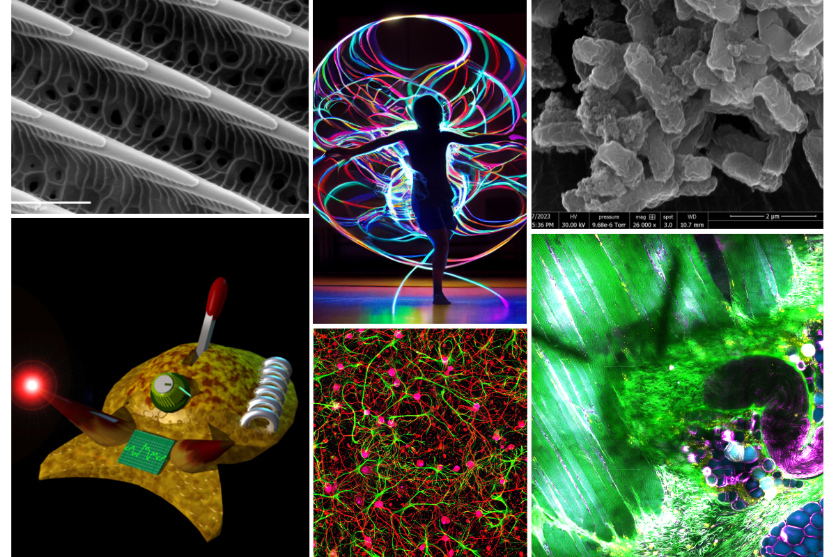
What do researchers look at all day? Perhaps a lush, green-and-purple rainforest of muscle fibers. Maybe a microscopic swallowtail scale. Sometimes, a computer-generated sea slug.
These perspectives and more are represented by the six winners of the Beckman Institute for Advanced Science and Technology's fifth annual research image contest. Each chosen image is associated with a different academic department on campus, and winners include students, postdocs, staff, and faculty members.
The contest is open to Beckman-affiliated researchers as well as non-Beckman affiliates on campus who use the institute's core facilities.
"It’s gratifying to see the diversity of Beckman's research reflected in these images," said Beckman Institute Director Nadya Mason. "This contest is just a small sample of the 40 academic units we represent, and it shows how many disciplines are impacted by the imaging facilities and capabilities here at Beckman. I love how the beauty and diversity of the science shines through."
A team of Beckman administrators, staff, and faculty members blind-judged the entries on their visual and research appeal. As in previous years, the winning images will be displayed for one year in the Beckman Director's Conference Room before finding permanent homes across the institute.
This year's winners are:
Graduate student category
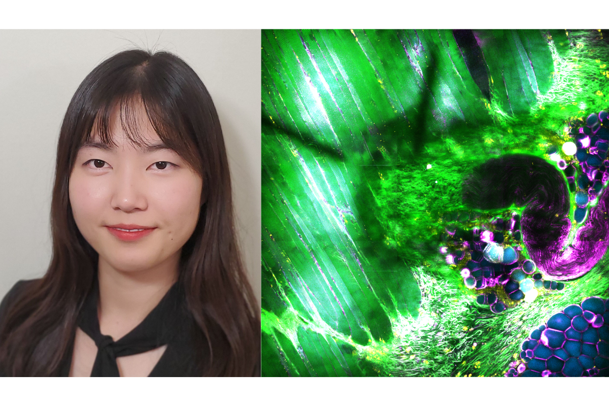 Jaena Park and her winning research image: Flora, Fibers, and Fluorescence.
Flora, Fibers, and Fluorescence
Resembling a rainforest canopy, mouse muscle fibers appear as elongated green structures, arranged in bundles with a distinct striated, or streaked, pattern. The fibers are separated by adipose tissue (cyan), nerve fibers (magenta), and other connective tissues, adding a burst of floral color to the scene. This image enables label-free visualization of the in-depth muscle tissue, offering a more accurate representation of its structure and composition without stains or dyes.
Jaena Park is a graduate student in the Department of Bioengineering. At the Beckman Institute, she collaborates with Professor Stephen Boppart in the Biophotonics Imaging Laboratory. This image was captured with a Simultaneous Label-free Autofluorescence-Multiharmonic, or SLAM, microscope, a laboratory-built microscope in the BIL.
Time Under Tension
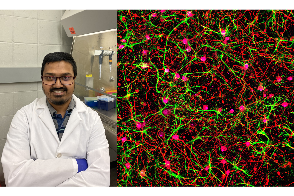 Md Saddam Hossain and his winning research image: Time Under Tension.
The human brain's 86 billion neurons strain under constant tension, enabling its neuronal network to generate enough mechanical force to lift a 2-pound weight. This image depicts a network of several hundred rat hippocampal neurons (red) surrounded by astrocytes (green). The force of the network pictured is on the order of 100 nanonewtons, which is roughly 5 million times weaker than the force needed to type on a computer.
Md Saddam Hossain is a graduate researcher in the Department of Mechanical Science and Engineering. At the Beckman Institute, Hossain collaborates with Professor Taher Saif within the Neurotechnology for Memory and Cognition Working Group. This image was generated with a laser confocal microscope in the Beckman Institute Microscopy Suite.
Staff and postdoc category
Tiger Scale
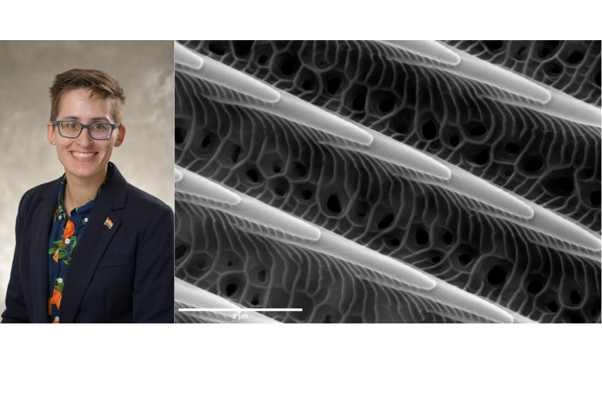 T. Josek and their winning research image: Tiger Scale.If you pick up a tiger swallowtail butterfly by its wings, you might notice what looks to be dust on your fingertips. That dust is made of small structures called scales. This image depicts a single tiger swallowtail scale magnified by 30,000 times. The scale’s lattice structure gives color to the butterfly’s wings; engineers have used it as a source of bioinspiration to add color to fabricated items.
T. Josek joined the Beckman Institute as a microscopist in June 2022. This image was generated with an environmental scanning electron microscope in the Beckman Institute Microscopy Suite.
Nanoplastics and Bacteria are in a Bind
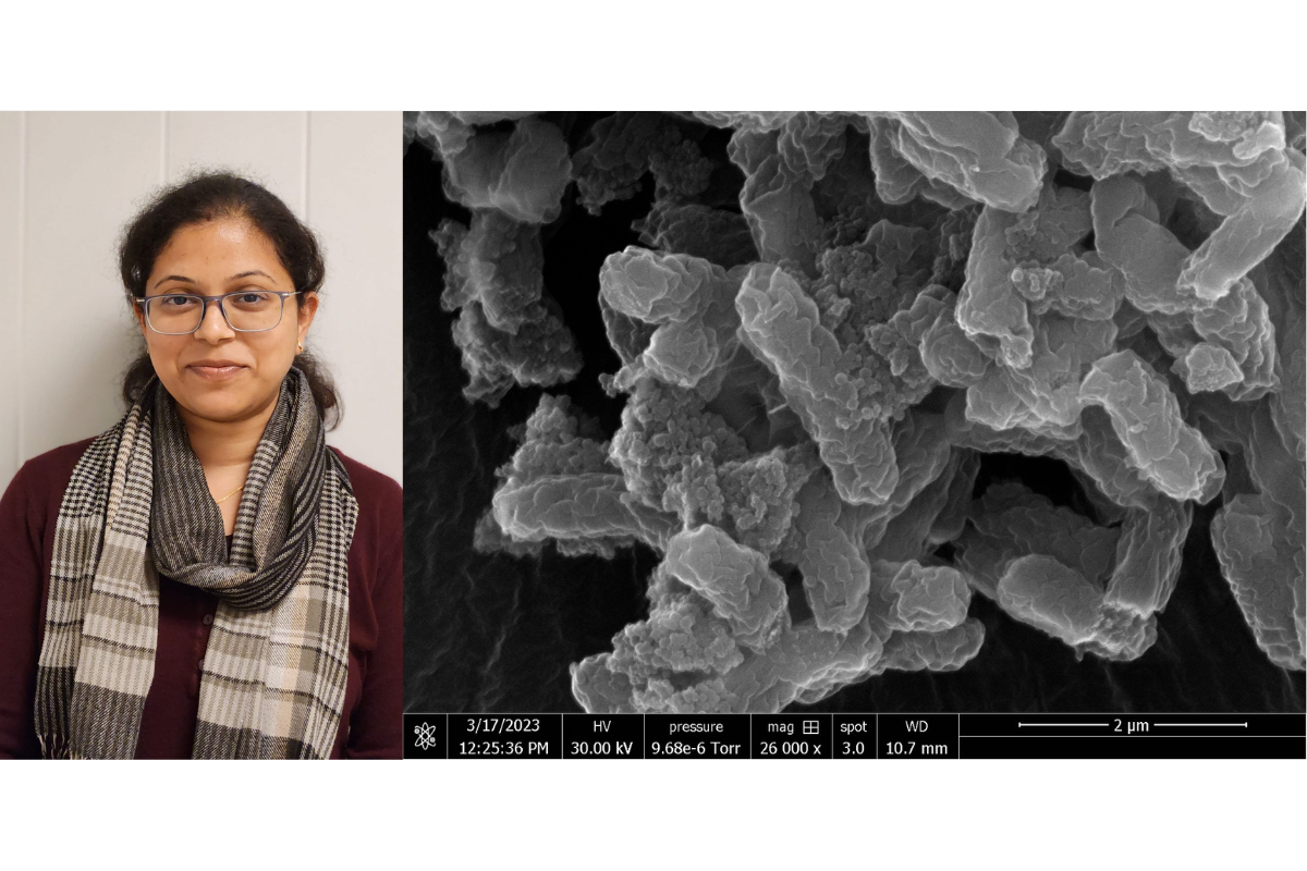 Jayashree Nath and her winning research image: Nanoplastics and Bacteria are in a Bind.
Nanoplastics are the emerging contaminants in our ecosystem. At less than 100 nanometers in size, the tiny plastics interact with bacteria by binding to the cell surface. This image depicts how clusters of polystyrene-based nanoplastics bind to the surface of the food-borne pathogen E. coli.
Jayashree Nath is a postdoctoral researcher collaborating with Professor Pratik Banerjee in the Department of Food Science and Human Nutrition. This image was generated with an environmental scanning electron microscope in the Beckman Institute Microscopy Suite.
Faculty category
Cyberslug
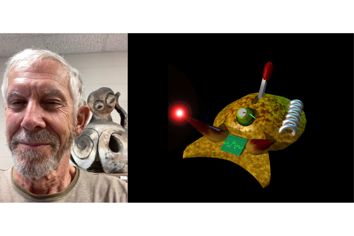 Rhanor Gillette and his winning research image: Cyberslug.
Cyberslug represents the simulation of decision-making circuits in the brain of the California sea slug, a predatory mollusk. When foraging, the slug weighs its hunger and experience against the qualities of potential prey; similarly, this agent-based software makes cost-benefit decisions. This image represents one of the first computational entities to simulate simple self-awareness.
Rhanor Gillette is a professor emeritus of molecular and integrative physiology and a researcher in the Neurotechnology for Memory and Cognition Working Group at the Beckman Institute. This image was generated by Mikhail Voloshin, who is an alumnus of Gillette’s lab.
Spectrum of Thought: Poi Spinner's Cognitive Dance
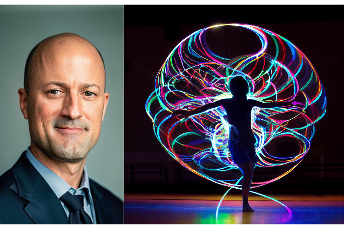 Sean Mullen and his winning research image: Spectrum of Thought: Poi Spinner's Cognitive Dance
This image blends physical flow movements with cognitive development. It depicts a luminous projection representing an infinite cerebral spin, illustrating the connective mind-body spark that fuels mental growth. This artistic representation portrayal accentuates the synergies among exercise, technology, and cognition for enhancing brain health.
Sean Mullen is an associate professor of kinesiology and community health and the director of the Exercise, Technology, and Cognition Lab. At the Beckman Institute, he conducts research within the Cognition, Lifespan Engagement, Aging, and Resilience Working Group. To create this image, a picture was taken of Mullen spinning LED lights attached to short tethers, a performance art called poi. Mullen used AI software to augment the image and generated a pixie to replace his silhouette.
To see more research images, please visit the Research Images page on our website or follow the Beckman Institute on social media.
Beckman Institute for Advanced Science and Technology