Article
High-powered magnetic fields, nuanced neuroimaging capabilities, the steady thudding of a scan in progress. An MRI is an MRI, right?
Not necessarily, according to the University of Illinois Urbana-Champaign and Carle Health. The institutions co-own and -operate a Siemens Healthineers MAGNETOM Terra 7 Tesla MRI scanner with transformative potential for research and clinical care.
Here’s everything you need to know about the first-of-its-kind collaboration.
1. What’s in the 7 Tesla’s name?
Strictly speaking, the MRI scanner in question is a Siemens Healthineers MAGNETOM Terra 7 Tesla MRI system. It’s a mouthful. Let’s break it down.
MRI is shorthand for magnetic resonance imaging. MRI scanners use a high-powered magnetic field to capture detailed visualizations of what’s going on inside our bodies.
Tesla is the scientific unit used to measure how strong magnets are. The strength of this particular magnet is 7 Tesla. For reference, a refrigerator magnet measures up at roughly 0.001 Tesla.
Siemens Healthineers, the healthcare division of global conglomerate Siemens GA, developed and distributed the MAGNETOM Terra model purchased by UIUC and Carle Health.
2. Whose scanner is it, anyway?
The 7 Tesla MRI scanner was purchased with equal funds from Carle Health and UIUC and represents a first-of-its-kind collaboration between the two.
Time spent running the machine is evenly divided, with each institution receiving 3.5 days out of the week to use it. Carle time is primarily used for clinical patients and clinical translational research, while Illinois time is primarily used for advanced neuroscience applications and the development of new technologies. Some projects are collaborative, with both institutions contributing time, tools, and expertise — Carle at the intersection of research and clinical diagnostics, and UIUC in the realm of cross-disciplinary study.
3. Where is the 7 Tesla located?
 The 7 Tesla MRI scanner co-owned and -operated by UIUC and Carle is located in the Carle Illinois Advanced Imaging Center in Urbana.
To create the most comfortable and convenient environment for patient care, the 7 Tesla scanner physically resides in the Carle Illinois Advanced Imaging Center in Carle MRI suites located at 611 W. Park St. in Urbana. In addition, researchers
at BIC and Carle’s Clinical Imaging Research Program are dedicated to the development and translation of imaging technology. Locating this jointly owned scanner in the clinical setting serves both institutions in their
effort to serve science and the community.
The 7 Tesla MRI scanner co-owned and -operated by UIUC and Carle is located in the Carle Illinois Advanced Imaging Center in Urbana.
To create the most comfortable and convenient environment for patient care, the 7 Tesla scanner physically resides in the Carle Illinois Advanced Imaging Center in Carle MRI suites located at 611 W. Park St. in Urbana. In addition, researchers
at BIC and Carle’s Clinical Imaging Research Program are dedicated to the development and translation of imaging technology. Locating this jointly owned scanner in the clinical setting serves both institutions in their
effort to serve science and the community.
4. How is the scanner used?
The MAGNETOM Terra 7 Tesla scanner has a unique dual-mode functionality, which means it can switch between clinical and research tasks to meet the specific needs of Carle and the university. This versatility is not something that’s seen every day, especially in a scanner of this strength, and it enables clinicians, researchers, and technicians to unite and face new challenges.
Currently, the Beckman Institute’s Biomedical Imaging Center, or BIC, operates three MRI scanners — two MAGNETOM Prisma 3 Tesla human whole-body MRI scanners and one Bruker 9.4 Tesla preclinical animal MRI scanner — and is responsible for the university’s engagement on the MAGNETOM Terra 7 Tesla scanner. Scientists use these scanners for biomedical, materials, agricultural, and fluid mechanics research.
5. Why is 7 Tesla technology exciting?
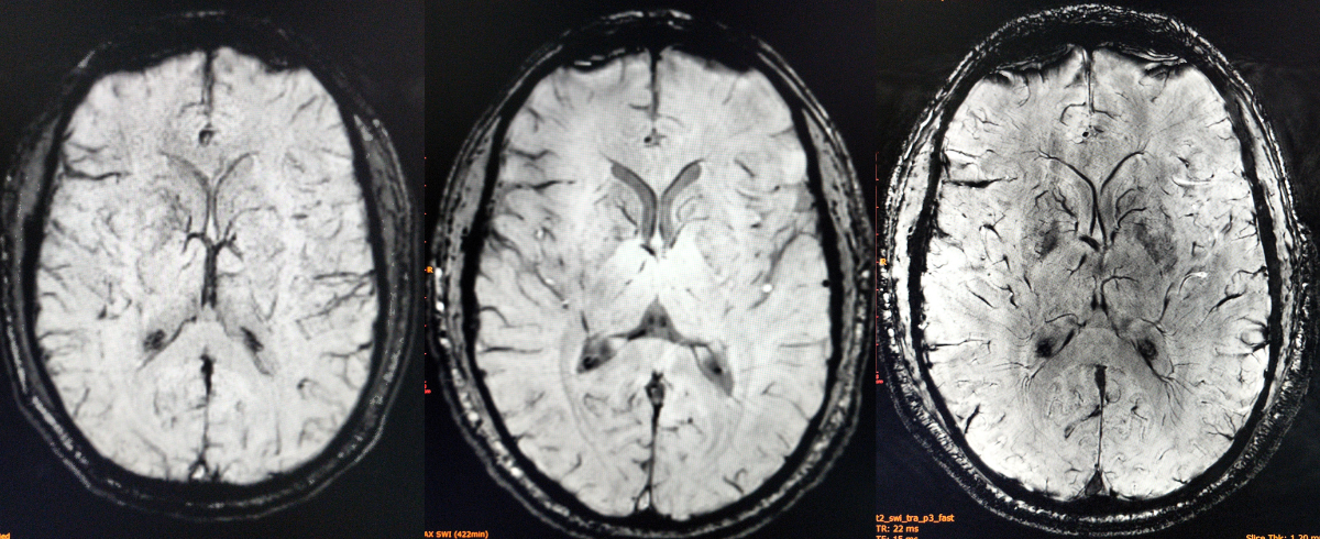 Three MRI scans of the same brain demonstrate the difference between 1.5 Tesla (left), 3 Tesla (center), and 7 Tesla (right) MRI. Credit: Carle Health.
If you’ve never been up close and personal with an MRI scanner — or if you’re accustomed to being inside the machine rather than in the control room — the differences between 1.5 Tesla, 3 Tesla, and 7 Tesla field strengths may seem subtle.
Far from it: images generated at 7 Tesla field strength aren’t just sharper than their lower-field counterparts — they’re transformative. The high level of detail allows researchers and clinicians to make observations that would not otherwise be possible.
These observations can be life-changing, and even lifesaving. For example, imaging the brain at 7 Tesla field strength can be used to definitively identify and diagnose early signs of focal cortical dysplasia, or epilepsy, a congenital condition that causes defects in the growth of brain tissue that become a source of seizures.
6. How can patients and research participants stay safe in the scanner?
It’s easy! MRI scans are perfectly safe for fully approved individuals. A common misconception is that MRI scans expose patients and research participants to radiation: they do not.
The MAGNETOM Terra 7 Tesla scanner has been approved by the Food and Drug Administration for human clinical assessments of brains and knees. In fact, the FDA considers magnetic fields up to and including 8 Tesla as a non-significant risk device for anyone older than one month. Other body parts can be safely scanned for research studies, but not yet for clinical interpretation of disease or injury.
Since a 7 Tesla MRI scanner is, at its core, a very strong magnet, standard safety precautions do apply. Anyone receiving an MRI (at any field strength) participates in extensive screening to confirm that they do not have metal implants or implanted devices that should not enter a strong magnetic field. Although some electronic implants are compatible with magnets of lower field strengths, 7 Tesla MRIs are relatively new to clinical settings and testing for specific types of implants is ongoing.
For more information about MRI safety and best practices, visit: https://www.fda.gov/radiation-emitting-products/mri-magnetic-resonance-imaging/what-patients-should-know-having-mri-exam
7. How can I get involved?
 If you're reading this article, there are opportunities for you to engage with the 7 Tesla MRI scanner.If you’re reading this article, there are opportunities for you to engage with the 7 Tesla MRI scanner. Here are a few ways to get started:
- Learn. Visit the Illinois MRI Exhibit at the Beckman Institute to learn more about the history of MRI technology in Champaign-Urbana.
- Participate. The Champaign-Urbana Population Study is underway and actively recruiting participants. Read more about the comprehensive brain and cognitive health study and how you can take part at https://cupopulationstudy.illinois.edu/.
- Get in touch. All campus research regarding the MAGNETOM Terra 7 Tesla scanner is processed through the Biomedical Imaging Center at the Beckman Institute. If you have questions about a university project proposal, please email BIC at bic@beckman.illinois.edu.
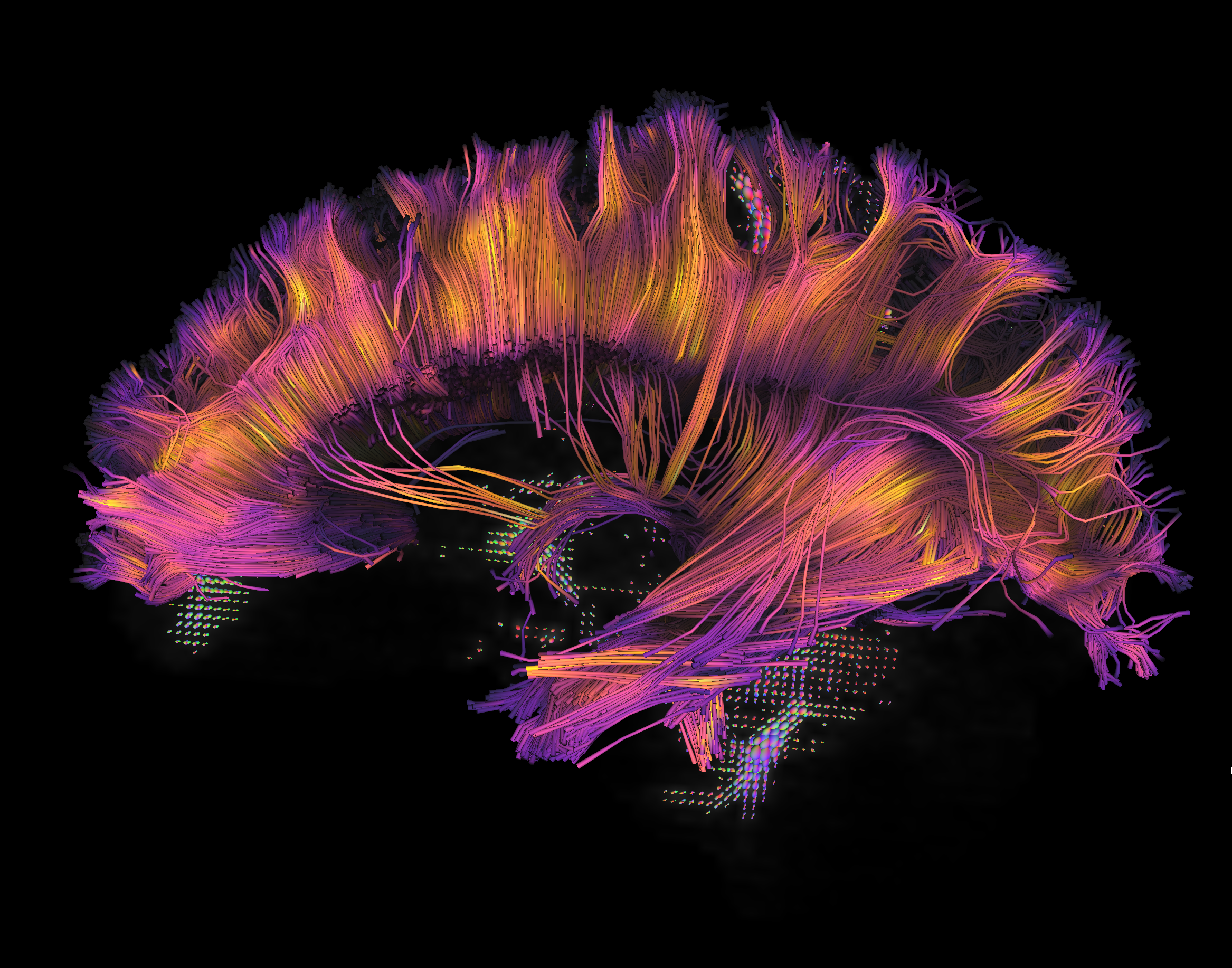
Banner image caption: Side view of a brain diffusion tractogram from a human participant showing fiber tracts of the cingulum and corpus callosum white matter bundles. Diffusion tractography uses brain imaging data from magnetic resonance imaging that is sensitive to the movement of water in the brain to estimate the directions of nerve fibers and how different parts of the brain are connected. Microstructural properties of the white matter can be measured along these nerve fibers, as displayed here by color along tracts changing from yellow in the more densely packed and myelinated center of the white matter bundles to purple as nerves branch out to connect to the cortex.
Beckman Institute for Advanced Science and Technology