Article
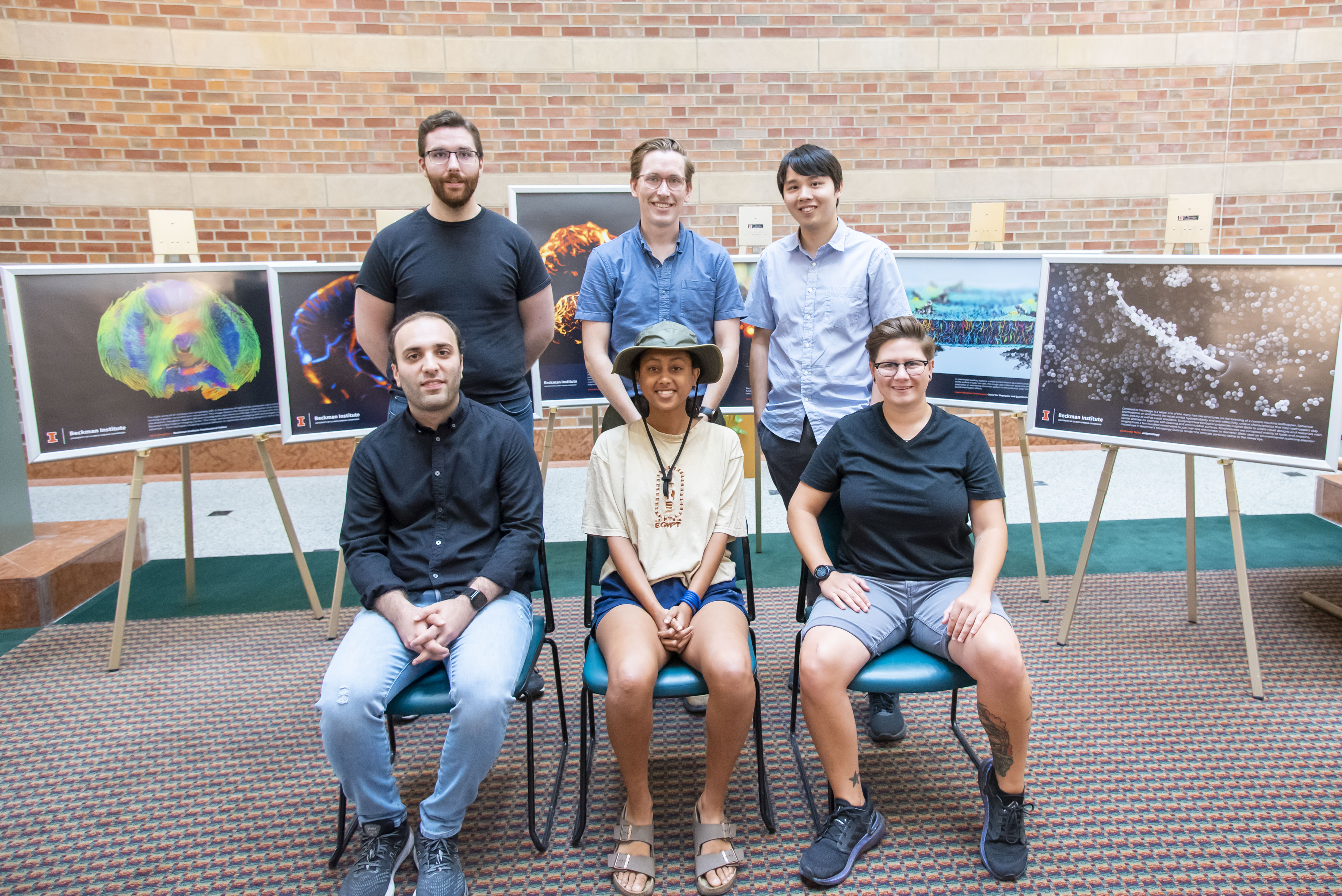 Six researchers were recognized in the 2022 Beckman Institute Research Image Contest: (top row) Matthew Lowerison, Noel Naughton, Shensheng Zhao; (bottom row) Sepehr Dehghani-Ghahnaviyeh, Alayt Issak, Elizabeth Bello.
Artwork adorns the halls of the Beckman Institute for Advanced Science and Technology, but it's not just for show — the images on the walls are evidence of the research taking place within them.
Six submissions from the 2022 Beckman Institute Research Image Contest are the latest to be framed and displayed in the Beckman Director's Conference Room. The winners include graduate students, postdoctoral researchers, and staff members from departments across campus.
“Sharing research images is one of the most appropriate, exciting ways to celebrate the institute’s shared imaging resources,” said Beckman Director Jeff Moore. “The idea that researchers from anywhere on campus can come here and use some of the best imaging technology in the world to advance their own research and see things that have literally never been visualized before — that’s Beckman’s bread and butter, and the winning images are a testament to that.”
In the tradition of the contest, which is now in its fourth year, the chosen images from last year will be hung in new locations throughout the building. Research images from 2022 and 2021 will also be displayed on easels in the Beckman Atrium throughout the month of August.
A team of Beckman administrators, staff, and faculty members blind-judged the entries on their visual and research appeal. This year's winners are:
Graduate student category
Winner: Shensheng Zhao, electrical and computer engineering
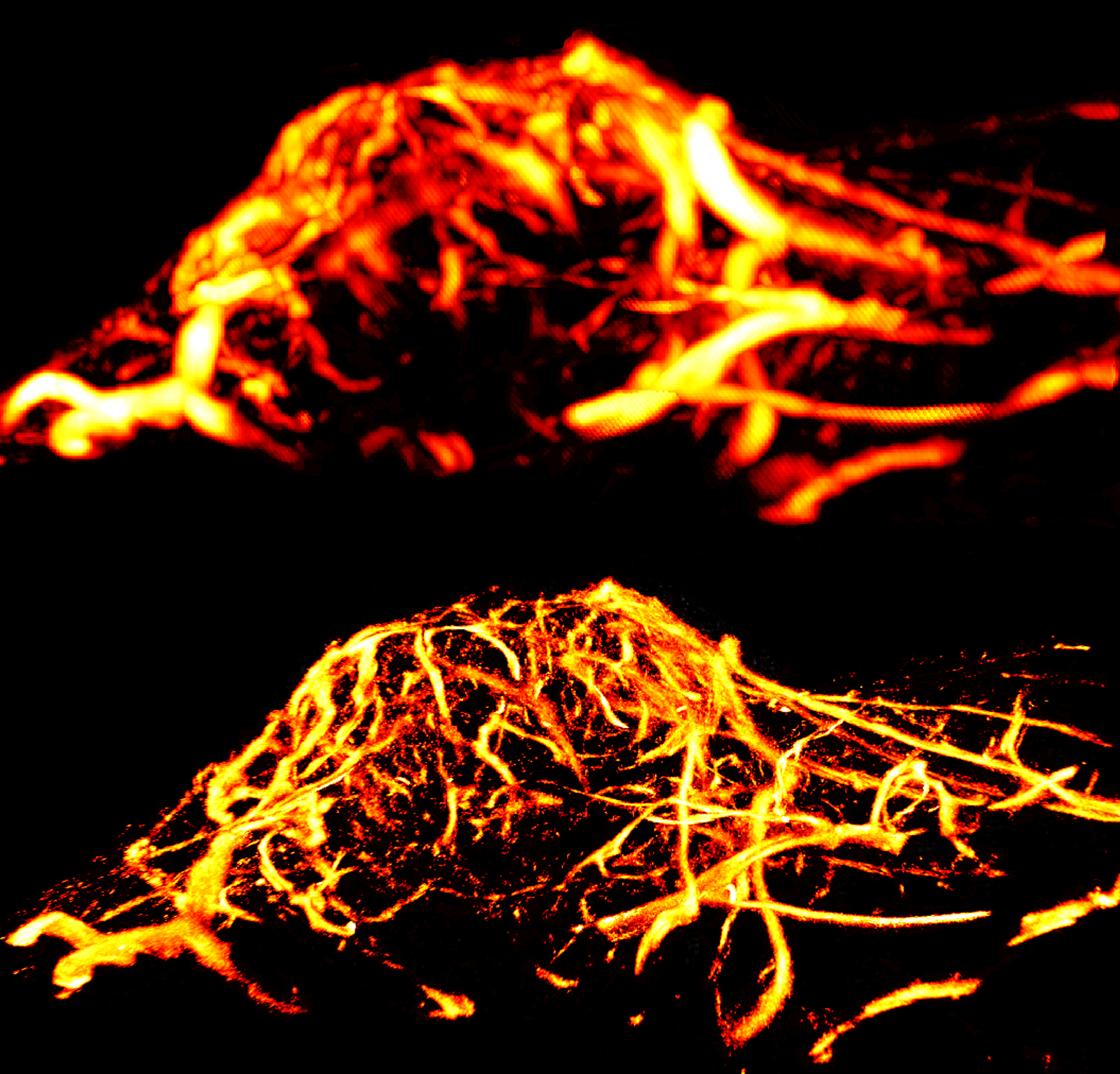 Shensheng Zhao submitted this winning image to the 2022 Beckman Institute Research Image Contest in the graduate student category.
Assessing a tumor's malignancy often requires looking at its microscopic network of blood vessels and veins. This image contrasts two ultrasound imaging techniques used to visualize tumor microvasculature in mice: power Doppler imaging (top) and the much finer ultrasound localization microscopy (bottom). Zhao's research involves augmenting ULM with machine learning to help make ultrasound imaging a more accurate diagnostic tool and advance preclinical cancer research.
Shensheng Zhao is a graduate student in the Department of Electrical and Computer Engineering. At Beckman, he collaborates with Professor Yun-Sheng Chen in the Extracellular Vesicle Imaging and Therapy Working Group.
Honorable mention: Elizabeth Bello, entomology
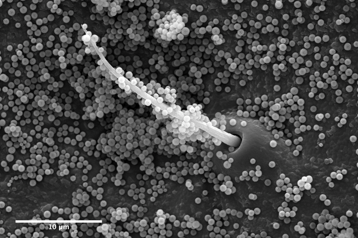 Elizabeth Bello's research image received an honorable mention in the 2022 Beckman Institute Research Image Contest in the graduate student category.
Centered in this image is a setae: one of the many hair-like structures on the forewing of a Curtara insularis leafhopper. Spherical brochosomes on and around the setae exhibit hydrophobicity and anti-reflectivity, unique characteristics that help leafhoppers stay clean and dry as well as evade predators. Studying these biological properties inspires Bello to create novel designs and materials with similar abilities; for example, self-cleaning and antimicrobial surfaces. Brochosomes are composed mainly of lipids and proteins, ranging from a few hundred nanometers to just over one micrometer in diameter. Bello used an environmental scanning electron microscope in Beckman's Microscopy Suite to render the intricate particles visible to the naked eye.
Elizabeth Bello is a graduate student in the Department of Entomology and a member of the Alleyne Bioinspiration Col-LAB-orative. She is a Beckman Institute Graduate Fellow and conducts research alongside the Schroeder Lab Group.
Honorable mention: Sepehr Dehghani-Ghahnaviyeh, Center for Biophysics and Quantitative Biology
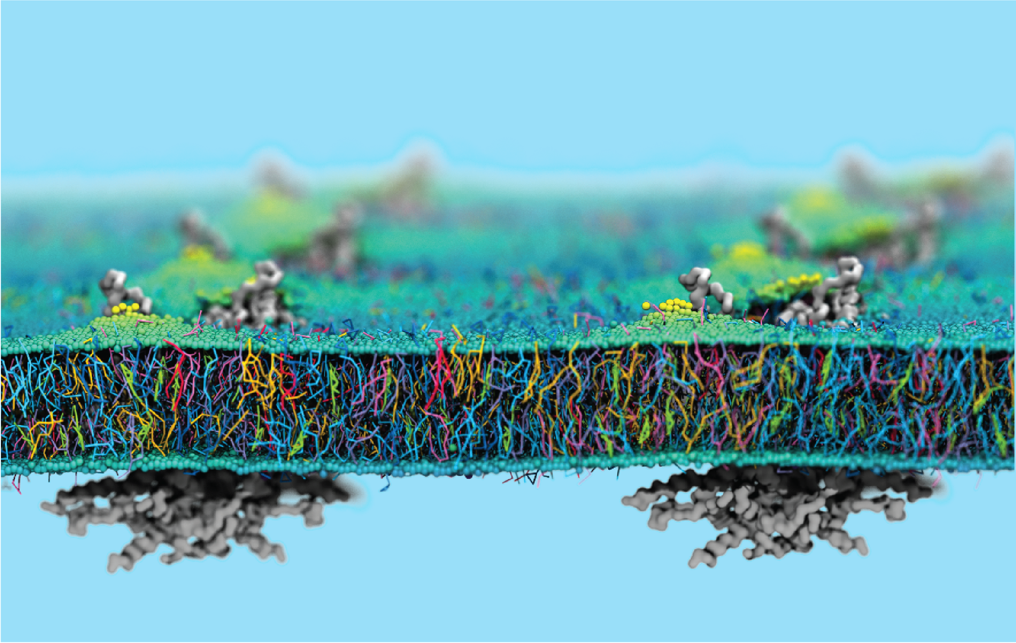 Sepehr Dehghani-Ghahnaviyeh's research image received an honorable mention in the 2022 Beckman Institute Research Image Contest in the graduate student category.
In mammalian auditory systems, a motor protein known as prestin functions as the sound amplifier. Prestin is located on the surface of outer hair cells with a high density. This image uses molecular dynamics simulations to show that prestin dimers (shown in grey) follow an astonishing placement pattern on the surface of outer hair cells (shown in rainbow colors) to maximize their sound amplification effects.
Sepehr Dehghani-Ghahnaviyeh is a graduate student in the Center for Biophysics and Quantitative Biology. At the Beckman Institute, he conducts research in the Theoretical and Computational Biophysics Group.
Staff/postdoctoral researcher category
Winner: Noel Naughton, Beckman Institute Postdoctoral Fellow
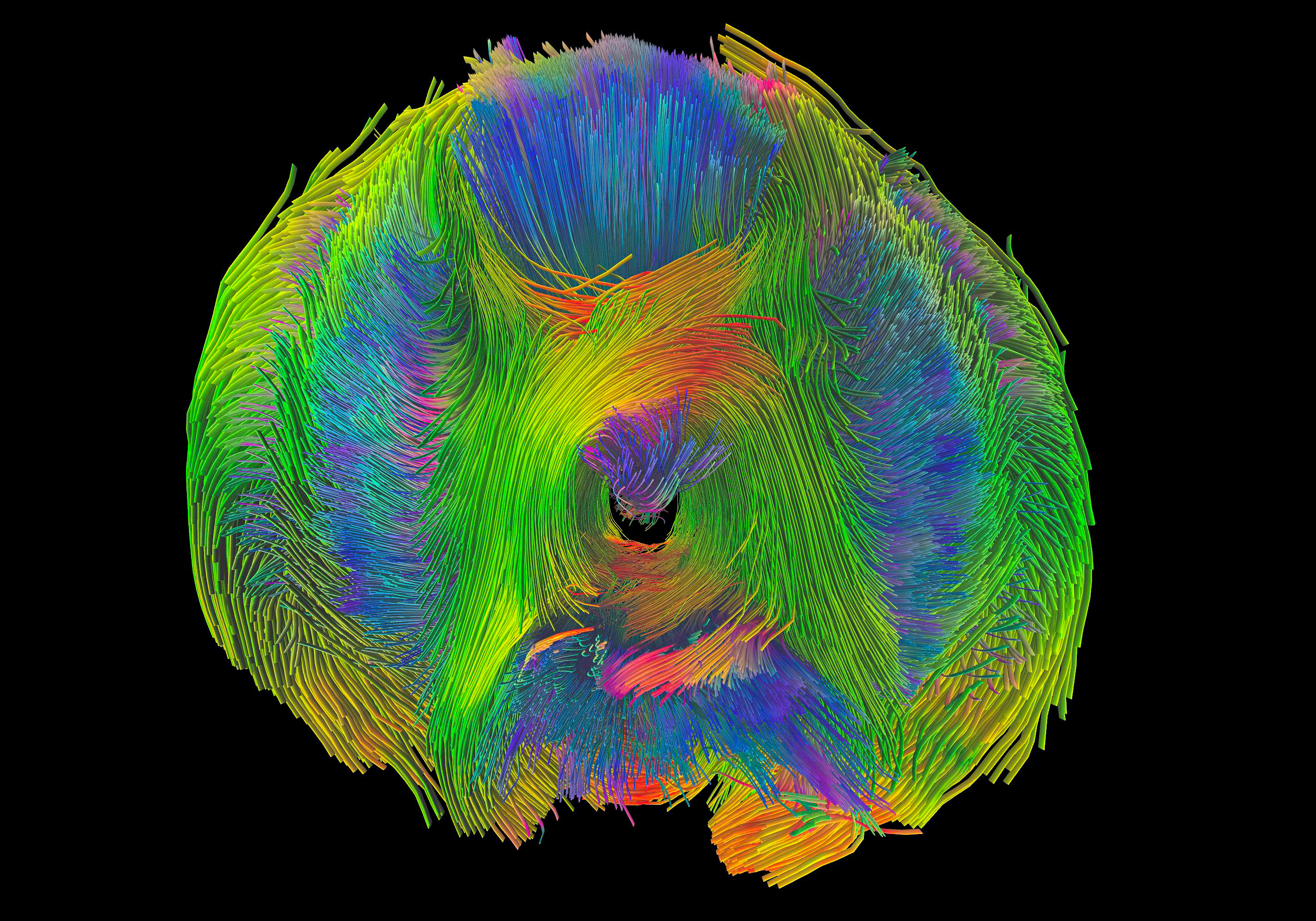 Noel Naughton submitted this winning image to the 2022 Beckman Institute Research Image Contest in the staff/postdoctoral researcher category.
The eight arms of an octopus are completely soft. In the absence of rigid bones, octopuses use a complex arrangement of muscles working together to control arm movement. This image depicts the muscular organization of an octopus arm in 3D.
To create this visualization, Naughton applied diffusion tractography algorithms to MRI data of an octopus arm acquired on the 9.4 Tesla scanner in Beckman's Biomedical Imaging Center. The image demonstrates how emerging MRI technologies can help researchers make new advances — from biologists studying octopus behavior to roboticists developing designs to mimic the octopus' abilities.
Noel Naughton is a Beckman Institute Postdoctoral Fellow conducting research in the Brain Connectivity and Networks Working Group.
Honorable mention: Matthew Lowerison, Beckman Institute Postdoctoral Fellow
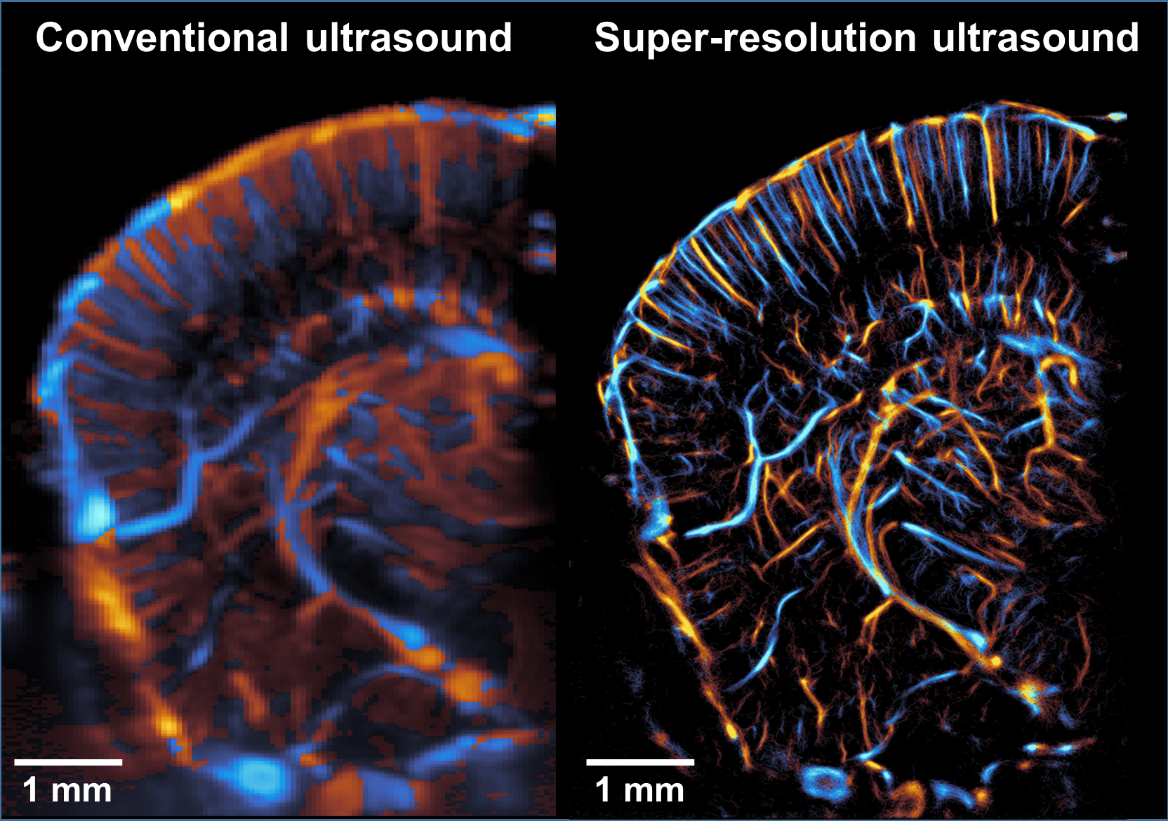 Matthew Lowerison's research image received an honorable mention in the 2022 Beckman Institute Research Image Contest in the staff/postdoctoral researcher category.
Two imaging techniques are contrasted in this ultrasound image of a mouse brain hemisphere: conventional ultrasound imaging (left); and super-resolution ultrasound imaging (right), a clinically translatable method developed by Lowerison and his colleagues to measure microvascular blood flow in the brain without compromising imaging depth. His team is applying this technique to better understand the relationships between the brain's vascular system, aging, and Alzheimer's disease, ultimately seeking to identify risk factors related to microvasculature that are associated with cognitive impairment.
Matthew Lowerison is a Beckman Institute Postdoctoral Fellow conducting research in collaboration with the Llano Lab and Song Lab.
Honorable mention: Alayt Issak, Coordinated Science Laboratory
 Alayt Issak's research image received an honorable mention in the 2022 Beckman Institute Research Image Contest in the staff/postdoctoral researcher category.
Electrical activity in fungi is depicted in striking 3D. Generated with artificial intelligence, this image challenges the fundamental limit of machines — creativity — and introduces a world where machines can co-create with artists.
Alayt Issak is a is a predoctoral fellow in the Coordinated Science Laboratory. She collaborates with Professor Lav Varshney in the Information and Intelligence Group.
To see more research images, please visit the Research Images page on our website or follow the Beckman Institute on social media.
Beckman Institute for Advanced Science and Technology