Article
This Q&A is part of the Grad Student Visionaries series, a spin-off of our Student Researcher Spotlight that highlights the state-of-the-art equipment housed in Beckman's Microscopy Suite and Visualization Laboratory and the graduate students who use it.
 Elizabeth BelloLeafhoppers, cicadas, and dragonflies…oh my! Elizabeth Bello is an Illinois graduate student in the Department of Entomology who’s extensively studied all three of these creepy crawlers and arrays of other insects as well. She is a researcher in the Alleyne Bioinspiration Co-LAB-orative (ABCLab) and works alongside Marianne Alleyne, an associate professor of entomology. Her research seeks to discover how new technologies can learn from biological adaptations found in nature.
Can you explain your research?
Broadly, I study bioinspired designs and materials, which is how we as humans can use adaptations found in the natural world to create new and innovative technologies. Currently, I am researching brochosome morphology, wettability, and the surface structure of leafhopper insects. These brochosomes and cuticular features give the leafhoppers unique properties such as hydrophobicity and anti-reflectivity, which likely aid in self-cleaning, camouflaging, and predator defenses.
What Microscopy Suite tool do you use the most, and why?
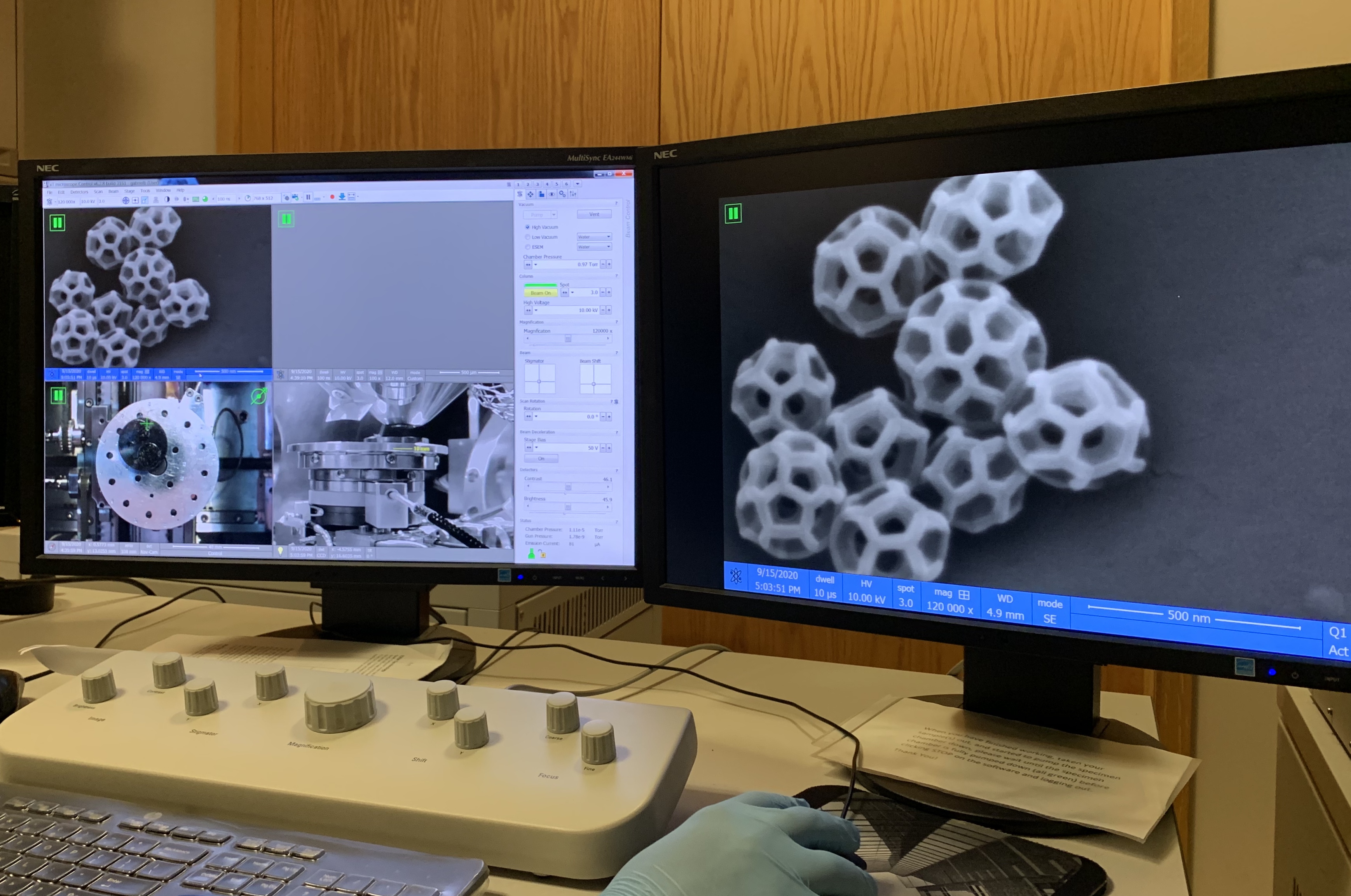 Imaging brochosomes at Beckman. Credit: Elizabeth Bello.For this research project, I've been using the environmental scanning electron microscope the most. The ESEM allows me to visualize and measure the brochosomes and topographical features found on various leafhopper species. I’m also exploring a SEM nanocondensation technique which will allow me to measure the wettability of leafhopper wings in situ with greater visual detail than a typical micro goniometer.
Can you share a technique that’s helpful to you on the ESEM?
To image brochosomes and the cuticle topographies of leafhoppers, I normally use the standard SEM mode with a low-voltage, high-vacuum setting. I typically work with about 10kV at a spot size of 3.0 and a working distance of 5-10mm depending on the sample. While the ESEM has a control panel with knobs to adjust settings like contrast, brightness, magnification, focus, and astigmatism, I prefer to use the mouse and keyboard shortcuts. By right-clicking the mouse and dragging to the left or right you can adjust the focus. By holding down the shift key, right-clicking, and dragging left/right and up/down, you can correct the astigmatism. As long as the sample is properly grounded, toggling back and forth between adjusting the focus and the astigmatism will result in the clearest image.
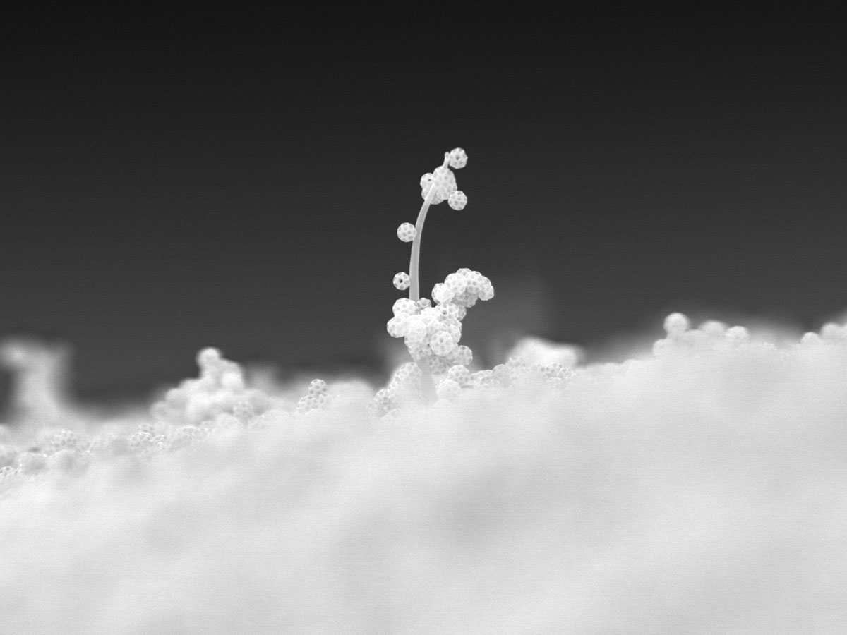 Leafhoppers can use camouflage to steer clear of predators thanks to brochosomes, miniscule spheres on the outer layer of the insect's body. Brochosomes are also hydrophobic, which helps keep leafhoppers dry. This image depicts brochosomes clinging to a leafhopper's wing setae, a bristly hair similar to a whisker. Credit: Elizabeth Bello.
My best advice in capturing clean, crisp images is to learn why an image doesn’t look right and what needs to be adjusted in order to improve it. If the image is oversaturated with white areas or it looks wavy and distorted, you can try adjusting the contrast. If that doesn’t work, it’s most likely an electron charging issue. The voltage can be lowered to reduce charging, but if that doesn’t resolve the issue, the sample probably needs to be prepared better to enhance grounding. If the image stretches while adjusting the focus, there is most likely an astigmatism which can be corrected. If the image still doesn’t look right, adjusting the lens alignment could result in better imaging. With time and practice, the quality of my images has definitely improved using these techniques.
Any words of wisdom for future visionaries?
I think my best piece of advice would be to always ask questions. Don’t be afraid of looking silly or that you’re asking a “dumb” question — there’s no such thing. The staff members in the Vis Lab all have tons of experience and are more than willing to help answer any questions you have and further your capabilities. It’s also wise to be patient with yourself. You’re still learning and it can take a while to get the hang of things. Just keep practicing and as your skills grow, your confidence will too. Best of luck and don’t be afraid to seek help!
For more information about Beckman Institute's Microscopy Suite, please visit: https://itg.beckman.illinois.edu/microscopy_suite
Beckman Institute for Advanced Science and Technology