Article
The Beckman Institute for Advanced Science and Technology has announced this year’s winners of its research image contest. They include undergraduate and graduate researchers, a research staff member, a faculty member, and two graduate students who have been researching the SARS-CoV-2 virus, which causes COVID-19. A team of Beckman administrators blind-judged the entries on their visual and research appeal.
“Every year, I’m amazed at the quality of both the research being conducted, and the beautiful images that result,” said Beckman Institute Director Jeff Moore. “As more people return to our building after long absences caused by COVID-19, I’m looking forward to seeing these images in person. They’re a great reminder of Beckman’s commitment to interdisciplinary, barrier-breaking research.”
This year’s special category on COVID-19-related research reflects the agility with which those researchers have examined and worked to understand the virus.
The posters highlighting last year’s winners will be distributed throughout Beckman.
This year's winners are:
 Shreyas Rajagopalan, undergraduate student category
Undergraduate student category
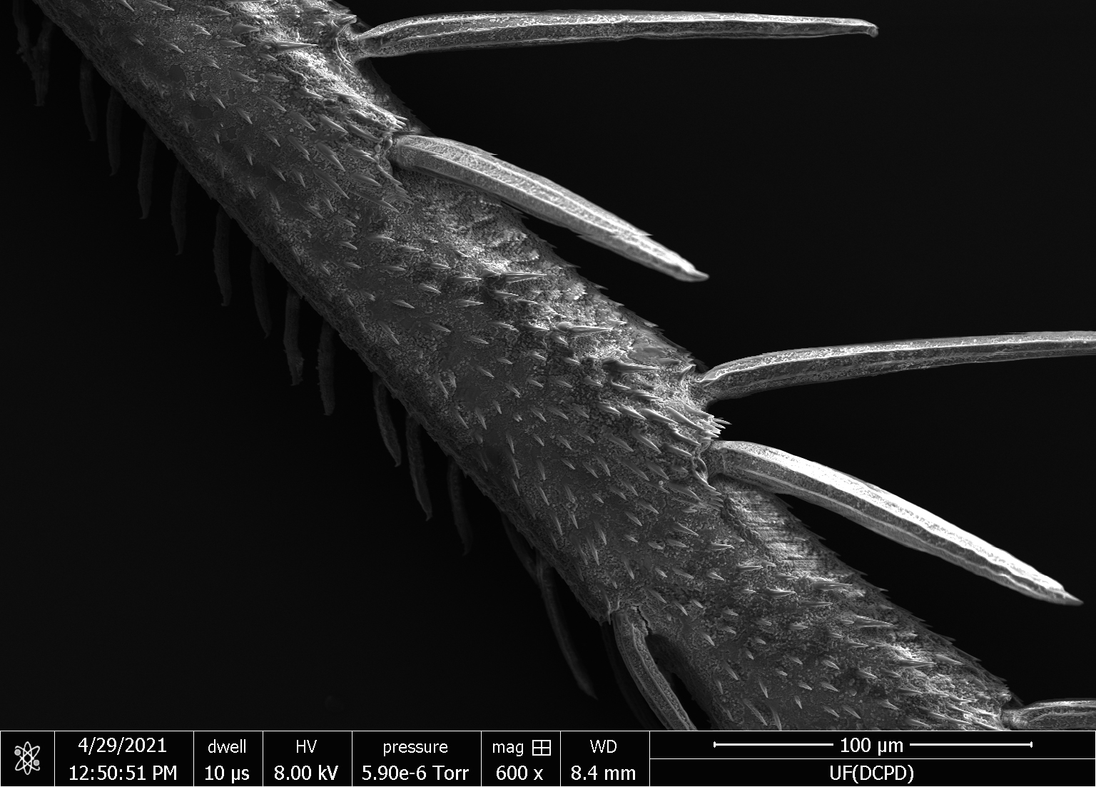
Shreyas Rajagopalan, Alleyne Bioinspiration Col-LAB-orative
This scanning electron microscope image of the tibia of the Emocasca leafhopper allows for a better understanding of its topography. The small spikes may represent sensory receptors that help the leafhopper detect vibrations. The image clearly illustrates how the brochosomes (the small dots) and topography are laid out. This helps our lab’s research because we can see how the topography may affect the number of brochosomes found on various bodily structures on leafhoppers.
Rajagopalan, a junior majoring in integrative biology honors and clinical psychology, is also a Beckman Institute Undergraduate Fellow.
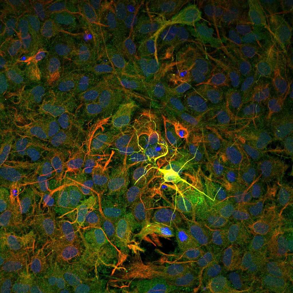 Ki Yun Lee, graduate student categoryGraduate student category
Ki Yun Lee, Saif Lab
A single neuron in yellow shines in front of an astrocyte. The astrocytes’ nuclei are green, and the somas and processes are red. This research examines the links between exercise and cognitive health. One of the ways to understand is to observe how astrocytes behave in neuronal environments. An astrocyte is a type of non-neuronal cell, and it provides energy and regulates factors that can enhance neuronal activities. Thanks to it, the neuron can thrive, function, and shine.
Lee is a graduate student from the Department of Mechanical Science and Engineering.
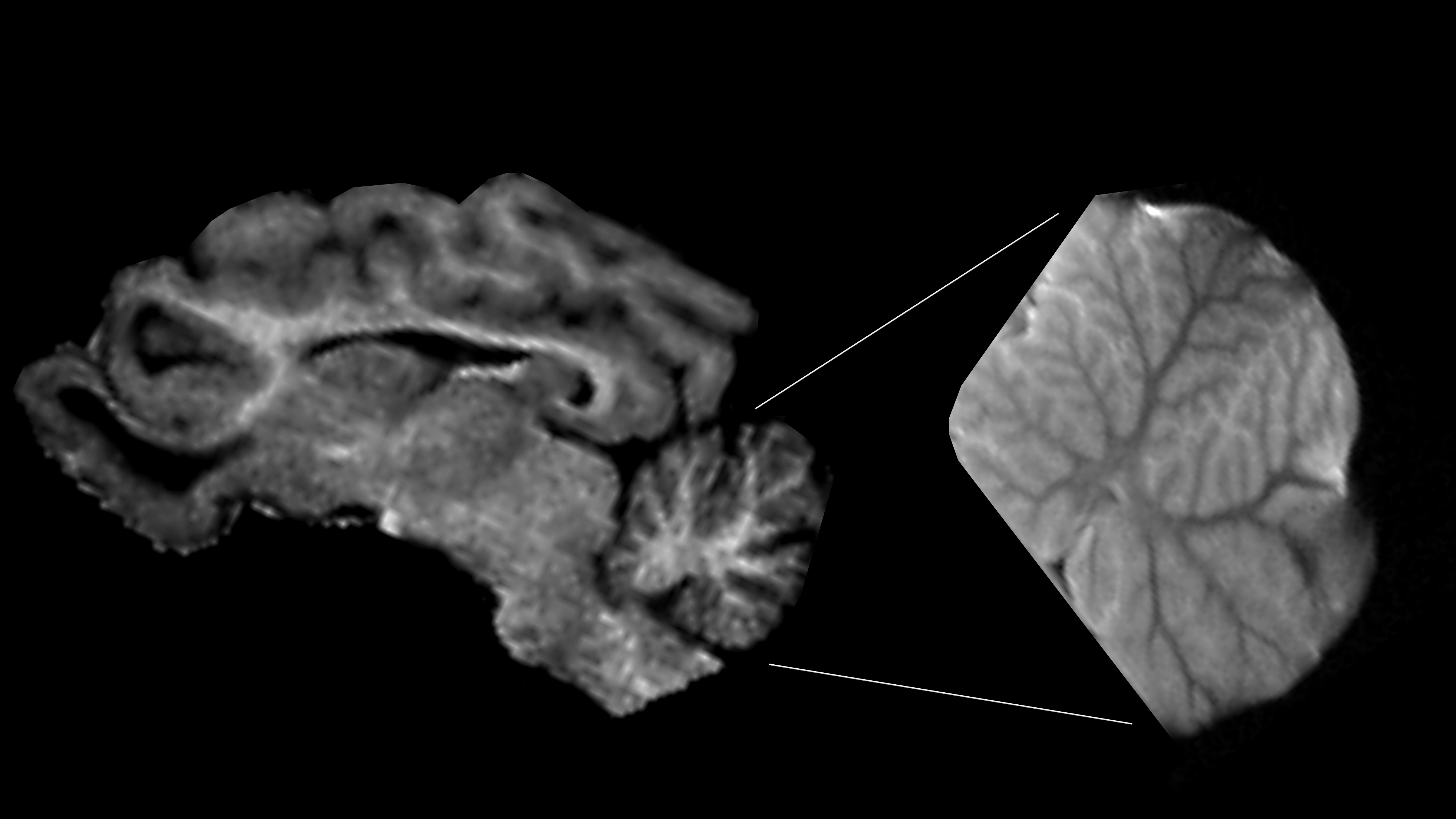 Ryan Larsen, postdoctoral and staff researcher categoryPostdoctoral and staff researcher category
Ryan Larsen, a researcher working with Andrew Steelman
MRI images taken of the brains of 4-week-old piglets. The left image was obtained using a 3 Tesla scanner and the right image was obtained using a 9.4 Tesla scanner. Because the magnetic field of the 9.4 Tesla scanner is over three times greater, the resolution is greatly improved. This enhanced resolution offers structural details of the cerebellum not possible to see at 3 Tesla. At the lower resolution, the folds (or lobules) of the cerebellar cortex appear to be thick and uniform. At the higher resolution, you can see each lobule contains many small folds, or folia, that give the cortex a beautiful leaf-like appearance. The 9.4 Tesla scanner has great potential for helping researchers determine the precise size and shape of many important brain regions. The measurements offer understanding into how the piglet’s diet affects the development of different parts of the brain. This knowledge will guide the development of human infant formulas that promote healthy brain development.
Other members of this research team include Shreyan Majumdar and Kwan-Jin Jung of Beckman’s Biomedical Imaging Center; Laurie Rund, Kayla Stanke, and Andrew Steelman of animal sciences; and Allison Louie of nutritional sciences.
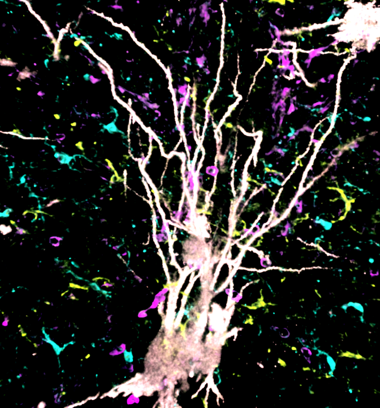 Makoto Inoue, faculty member categoryFaculty member category
Makoto Inoue, Department of Comparative Biosciences
A Golgi-Cox stained neuron in white is shown as it interacts with infiltrated T cells in purple, astrocytes in yellow, and microglia in cyan in a brain affected by murine autoimmune disease. This image was captured using the recently discovered confocal reflection super-resolution technique to visualize both metal-based Golgi-Cox neuron staining and immunofluorescence-tagged cells. Using this new technique, we can conduct precise and improved three-dimensional visualization and analysis. We can also identify the importance of interactions between neuron and peripheral immune cells migrated by the central nervous system’s resident glia cells (microglia and astrocyte) in the development of neurodegeneration under autoimmune disease conditions.
As our lab studies neuroimmunology, we are interested in how peripheral immune cells and the central nervous system’s neuronal plasticity affect one another in the presence of autoimmune, infectious, and neurodegenerative diseases. We’re also studying the effect of environmental and biological factors on such neuroimmune communications. The study using this new technique is creating new ideas in our field and led to publications in high-impact journals such as PNAS and Nature Communications. This knowledge of neuroimmune interactions will have an impact on many fields such as immunology, neurology, pathology, and psychiatry.
Rachel Khaw, a graduate student in the Neuroscience Program, collected this data, which was funded by NIH5R01AI136999.
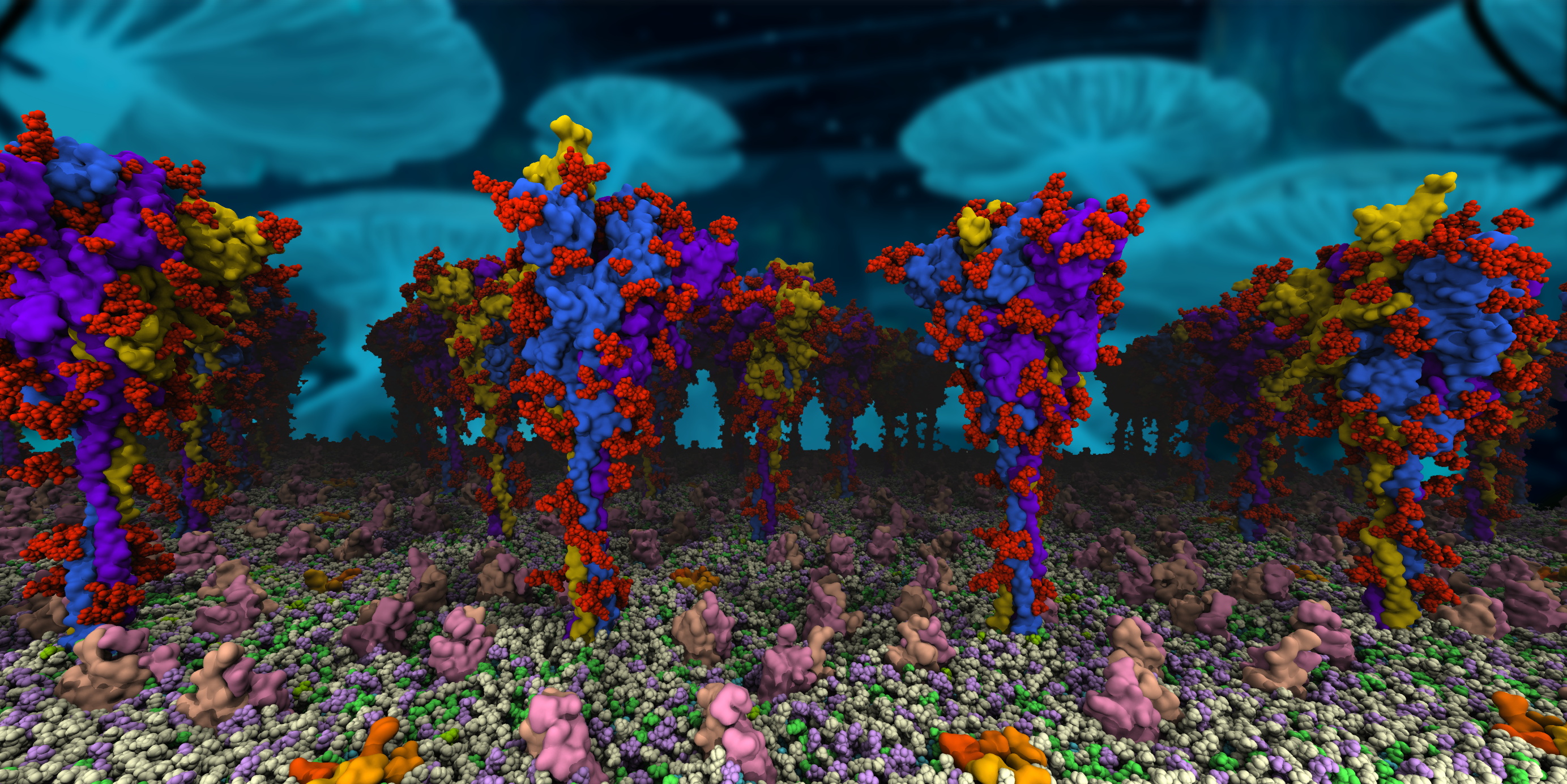 Charles Chen, special COVID-19 research categorySpecial COVID-19 research category
Charles Chen, Theoretical and Computational Biophysics Group
SARS-CoV-2 fuses to and enters human cells upon binding its spike proteins to human receptors. Our simulation model captures a realistic representation of spike proteins and their dynamics on the surface of the coronavirus. We rendered this image with Visual Molecular Dynamics, a program for displaying, animating, and analyzing large biomolecular systems using 3D graphics and built-in scripting. The Theoretical and Computational Biophysics Group at Beckman writes and distributes VMD.
Chen is a graduate student in the Center for Biophysics and Quantitative Biology.
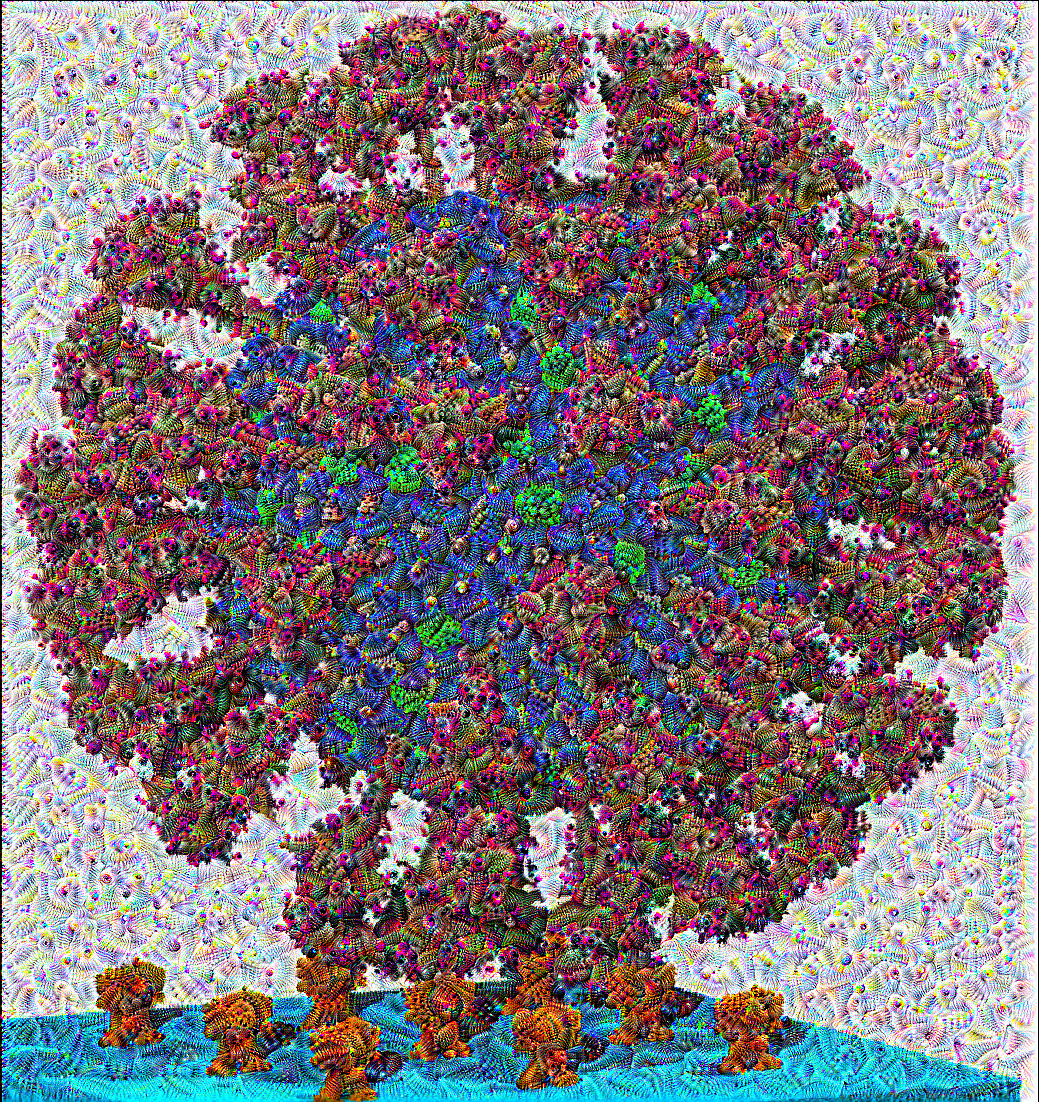 Hyun Park, special COVID-19 research categoryHyun Park, Theoretical and Computational Biophysics Group
This is a SARS-CoV-2 virus particle (the center object), near orange ACE2 human receptor proteins embedded on a cyan membrane. As part of our molecular dynamics research on the SARS-CoV-2 virus, the virus team in Professor Tajkhorshid’s group assembled a full-atom SARS-CoV-2 virus particle using Visual Molecular Dynamics software.
The VMD-generated image of SARS-CoV-2 has been transformed by a deep learning method called DeepDream (available on Google Tensorflow), which generates a hazy, mythical, and dream-like distortion of the original SARS-CoV-2 image with images of eyes (little black dots) drawn all over the original image. (The eyes in this case are mostly on magenta-colored glycans.) DeepDream captures the eyes feature it learned from animal images while training, and overlays them on top of any image a user inserts.
Just as Argus, Greek mythology’s all-seeing giant, watches vigilantly for enemies, the SARS-CoV-2 virus is a scary entity, endlessly seeking opportunities to attack humans with its many spike proteins that can sense human ACE2 receptors.
Park is a graduate student in the Center for Biophysics and Quantitative Biology.
Beckman Institute for Advanced Science and Technology