Article
Scientists at the Beckman Institute for Advanced Science and Technology recently showed off their research through Beckman Research Image Contest.
This year, the contest features four winners in the following categories: undergraduate students, graduate students, postdoctoral researcher, and faculty. The four framed images are being featured in the Beckman director’s conference room. Last year’s winning images will be hung throughout the Beckman’s halls.
“Researchers at the Beckman Institute use our state-of-the-art tools to work together across disciplines and break new barriers,” said Jeff Moore, the director of the Beckman Institute and an Ikenberry Endowed Chair in the Department of Chemistry. “These images show that research is not only important, but also visually beautiful. I continue to be amazed and inspired by the entries in the Beckman Research Image Contest.”
The winners are:
Undergraduate student category
Rachel Tham, the Minjoo Larry Lee Group
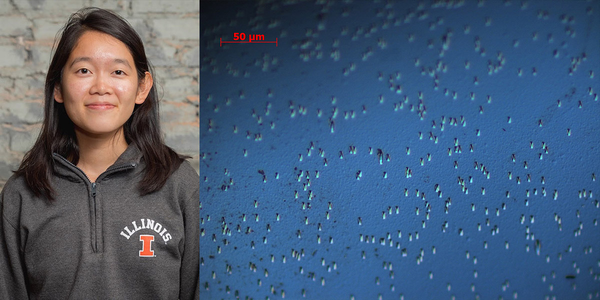 Multijunction solar cells could significantly increase the efficiency of solar power. However, lattice mismatch between the device layers can lead to defects called threading dislocations that decrease their efficiency. Tham's research, conducted with graduate student Ryan Hool, works to better understand why and how these dislocations occur to make multijunction solar cells more efficient. This image is a Nomarski (also called differential interference contrast) image of a beryllium-doped gallium phosphide (GaP) on GaP sample, after defect selective etching, and it was imaged with the Beckman Institute's inspection microscope at 50 times magnification. DSE reveals the location of threading dislocations as etch pits in the sample, and the number of etch pits present can then be used to calculate the sample's threading dislocation density.
Graduate student category
Marley Dewey, the Harley Research Lab
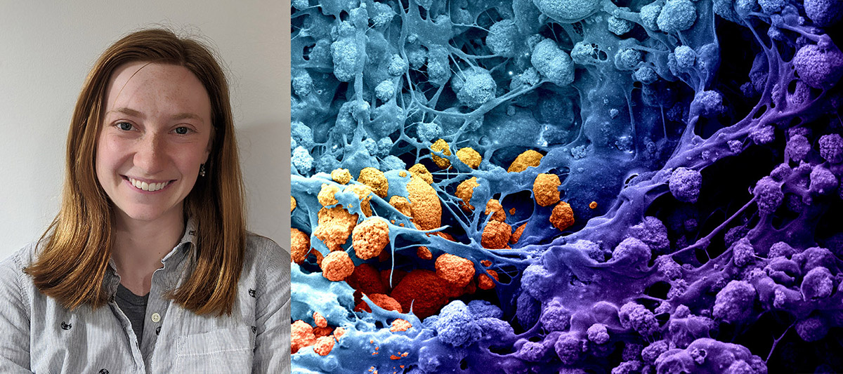 This is a mineral and polymer galaxy. This specific polymer contains calcium phosphate mineral and was 3D printed in order to repair damaged bone. Dewey’s role involves taking these 3D-printed polymers and combining them with collagen-based biomaterials in order to regenerate large missing portions of bone from the skull and jaw.
Postdoctoral and staff researcher category
Mark Levenstein, the Wagoner Johnson Applied Biomaterials and Biomechanics Lab 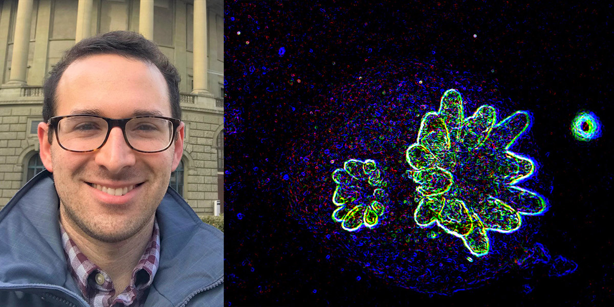
Optical micrograph of a baby Acropora palmata coral polyp inverted and modified for enhanced contrast of the newly formed tentacles. The polyp is shown growing on a novel carbonate reef restoration substrate, which hopefully will increase the settlement and survival of juvenile corals into adulthood.
Faculty member category
Brad Sutton, Magnetic Resonance Functional Imaging Lab
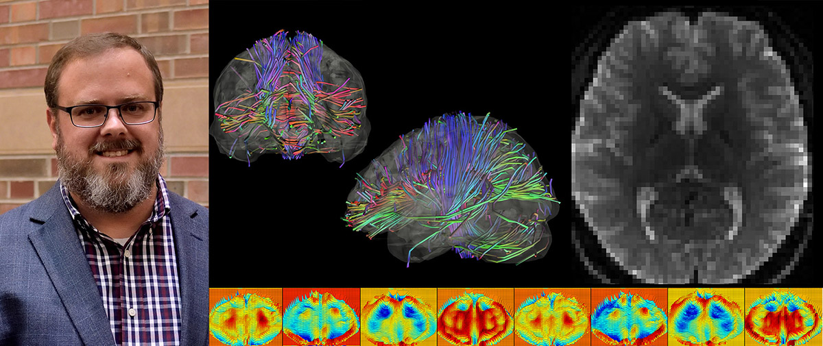 Two techniques in magnetic resonance imaging enable researchers to see highly sensitive information about the structure of brain tissue: diffusion tensor imaging and magnetic resonance elastography. DTI looks at the cabling in the brain. These white matter fiber pathways transmit information from one part of the brain to another. MRE looks at the mechanical properties of the brain tissue, including stiffness. It provides information about the interconnections and complexity of cells in the brain. In order to make a clinically feasible protocol and save time, the Sutton Group and Carle Foundation Hospital-Beckman Institute Postdoctoral Fellow Aaron Anderson, in collaboration with professors Dieter Klatt and Richard Magin at the University of Illinois at Chicago, developed and implemented a DTI-MRE sequence that acquires both DTI and MRE data at the same time. This data was collected on the Biomedical Imaging Center 3 Tesla Prisma MRI system. Grant funding: NIH/NIBIB 5R21EB026238-02.
Beckman Institute for Advanced Science and Technology