Article
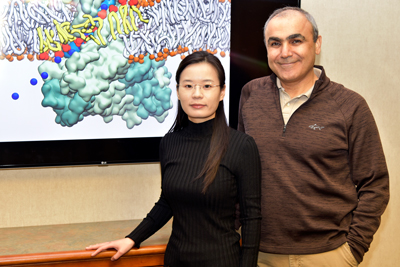
Members of the NIH Center for Macromolecular Modeling and Bioinformatics at the Beckman Institute recently published a paper that uses computational microscopy to visualize how lipids can influence the structure and function of protein channels in cells.
All living cells are surrounded by a membrane that is made of phospholipids. These molecules have a hydrophilic head group that is charged and faces outward, and long, uncharged, hydrophobic tails that reside in the core of the membrane.
Among this sea of phospholipids, there are proteins that span the membrane and mediate transport of materials such as ions and nutrients from one side to another. Lipid molecules not only provide the medium for membrane proteins, they also regulate their activity. However, experimentally, studying lipid and protein interactions is extremely challenging due to the fluid nature of lipids. Therefore, the researchers used computational methods to study these interactions.
Interestingly, a class of membrane proteins, known as lipid scramblases, are involved in transporting phospholipids from one side of the membrane to the other. Scramblases are special because the lipid-transporting activity of these proteins is affected by specific lipids in the membrane; proteins that affect the lipids and are at the same time affected by lipids.
The paper “A network of phosphatidylinositol 4,5-bisphosphate binding sites regulates gating of the Ca2+-activated Cl−channel ANO1 (TMEM16A)” looks at one such lipid-protein interaction and was published in the Proceedings of the National Academy of Sciences.
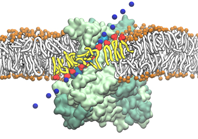
Usually the membrane proteins have hydrophobic groups that sit in the environment of the greasy phospholipid tails. However, the protein channel studied in the paper is a little different. “The interesting thing about this protein is that there is a hydrophilic pathway on the surface of the protein in the middle of the membrane. In our simulations, we see water fill up this cavity and the phospholipid heads use this pathway to move from one side of the membrane to the other,” said Tao Jiang, a postdoctoral researcher in the Tajkhorshid group.
“The tails are happy facing the greasy core of the membrane and the head just slides through like a credit card swipe,” said Emad Tajkhorshid, a professor of biochemistry and the leader of the Theoretical and Computational Biophysics Group at Beckman. “This is a very close, two-way interaction between lipids and proteins where some lipid works with the protein to go to the other side while other lipids control the channel’s activity.”
The arrangement of the protein, which is controlled by calcium ions, determines whether it allows the lipids to go through. “The release of calcium from inside the cell makes the protein open and the pathway can be used by lipids to move across. The lipids can also work with the protein to form a pathway for ions to travel,” Tajkhorshid said.
“These simulations are difficult to achieve as they have to capture very slow molecular processes. We need to make carefully crafted protocols to reproduce the natural motion and function of these molecules,” Tajkhorshid said.
“Before our simulations, people had no idea about the molecular details of how the lipids traveled across the membrane. Using simulations, we can see the cavity that forms a pathway for the lipids to go through, and how the lipids can also bind to the cavity and make a pathway for ions to go through,” Jiang said.
“We tell our collaborators at Emory (Dr. Criss Hertzell) where to make the mutations on the proteins and they strongly validate our results through experiments,” Tajkhorshid said. “We have many more collaborators who would love to know how the lipids affect their protein’s function, and computational simulation is currently the only way to visualize such phenomena.”
Beckman Institute for Advanced Science and Technology