Article
On the battlefield, soldiers usually wear protective gear that covers most of their bodies, but in many cases the Kevlar helmets that soldiers wear cannot fully protect the face, head, and neck. A 2013 article in the journal Military Medicine reported on the increase in combat-related head, face, and neck injuries among service personnel wearing combat body armor.
With funding from the U.S. Army, researchers at the University of Illinois are looking for ways to repair complicated skull injuries with biomaterials—substances that can interact with or guide the body’s natural healing processes—instead of using artificial materials like titanium plates or grafting bone from other areas onto the head.
“The defects of the skull typically in battle injuries are very strange in shape, so we want to be able to design a scaffold or composite that can fit that exact shape for them and then it would regenerate their own bone,” said Marley Dewey, a doctoral student working in the lab of Brendan Harley, an associate professor of chemical and biomolecular engineering.
“These severe craniomaxillofacial (CMF) injuries present a series of unmet clinical challenges to improved healing,” Harley said. “And while tissue engineering approaches suggest the potential to improve healing, we must first address a series of bottlenecks. Implants must balance very real considerations regarding strength and load bearing, the need to fit complex defects unique to each patient, and biotransport of nutrients during healing.”
According to Harley, the project seeks to pioneer approaches to integrate a bioactive collagen biomaterial that includes micro-scale porosity with a strong reinforcement frame generated via 3D-printing with macro-scale porosity.
“Such composites are common in other areas of engineering, such as reinforced concrete,” said Harley, “but not yet in tissue engineering, presenting us with some unique opportunities to advance our field.”
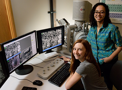
Dewey and her colleague Aleczandria Tiffany, also a Ph.D. candidate, are using mineralized collagen to regenerate bone. The tools in Beckman’s Microscopy Suite help them see if they are getting the desired results.
Dewey examines how 3D-printed biomaterials can be used to reinforce the mineralized collagen scaffold, which isn’t as strong as regular bone. The goal is to create a composite composed of biodegradable materials that break down over time after bone cells have started to proliferate. Deciding on the proper amount of scaffolding material can also be tricky. Dewey uses the environmental scanning electron microscope (ESEM) and the micro x-ray computed tomography (microCT) system in Beckman’s Microscopy Suite to examine cellular response on the composites.
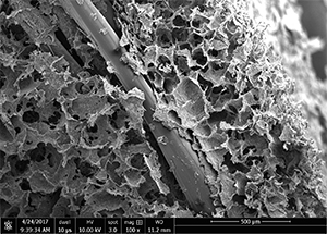
“I am looking for changes in structure and mineralization, but also to see that the 3D-printed materials are fully integrated into the mineralized collagen biomaterial, because gaps between them wouldn’t be ideal,” Dewey said.
“I also use microCT to see what the mineral content is. We have a program that a previous lab member developed using the microCT image files so you can quantify where the mineral is and how much is present.”
In addition, she uses the ESEM to examine the integration of anti-inflammatory agents incorporated into the collagen to combat bacterial infections and inflammatory responses that could be detrimental to soldiers experiencing a blast injury to the jaw on a battlefield, where it’s likely to be unhygienic.
Tiffany examines how incorporating growth factors—such as vascular endothelial growth factor and minerals such as zinc—work in helping an individual’s own bone grow within the scaffold.
“The scaffold is really porous,” Tiffany said. “Ideally the patient’s stem cells, already present at the wound site after injury, would migrate into it. So while Marley wants to add mechanical support, I want to add the growth factors so that those stem cells can be recruited into the material to accelerate bone regeneration.”
Zinc is incorporated into the scaffolds to promote mineralization and induce differentiation of stem cells. Using the ESEM allows Tiffany to see how zinc affects the microstructure within the scaffolds.
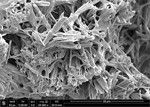
“Originally, I saw that when I added zinc there were no negative effects on overall pore structure, but then I could see that there was a definite difference in mineral microstructures with the addition of zinc,” Tiffany said. “We usually see ‘classic’ brushite plates, but with the addition of the zinc it’s very different. So I’ve added different concentrations of zinc to see how the mineral deposits change.”
These differences in microstructure may alter how stem cells generating bone interact with the scaffold. A cell’s microenvironment dictates its behavior, and this is something Tiffany hopes to explore more fully.
“Ideally I would want to see a cellular response to the zinc,” Tiffany said. “Hopefully with the addition of zinc we would see an increase in differentiation into osteoblasts and the expression of bone-related genes.” She hopes to use critical point drying at Beckman to examine how the cell’s morphology changes in her various scaffold types.
Both Dewey and Tiffany appreciate the one-on-one support from the Microscopy Suite staff.
“I had no knowledge of microCT or ESEM before I came here,” Dewey said. “Because my background was in chemical engineering, we didn’t use any microscopy tools. I didn’t even know what ESEM was.”
“It’s obvious that both Marley and Alec are power hitters,” said Scott Robinson, manager of the Microscopy Suite. “They are really engaged in their work and doing a great job; and we’re very happy to have them using the suite.”
Dewey thinks that the Microscopy Suite staff’s assistance has helped her produce great images useful in publications and presentations.
Tiffany, likewise, hadn’t had experience with the microscopes and credits Cate Wallace, Leilei Yin, and Robinson with providing their expertise to help the project along.
“We want our users to get the best possible images out of the various microscopes, and if some samples are tricky, making it hard to consistently pull up those killer images, we might check in on them more often,” said Robinson. “People working in bio and in biomaterials can face more difficulties in imaging their samples than straight materials people, and sometimes they can use more help. We have years of experience to offer them.”
“Now it’s at the point where Scott will come in and say ‘beautiful picture, you don’t need my help,’” Tiffany said. “But he still comes in almost every time I’m in there, just to tell me that the images are beautiful.”
Beckman Institute for Advanced Science and Technology