Article
Jonathan Sweedler, professor of chemistry, Martha Gillette, professor of cell and developmental biology, and Rohit Bhargava, professor of bioengineering, head up the project titled “BRAIN Initiative: Integrated Multimodal Analysis of Cell- and Circuit-Specific Activity using Mass Spectrometry Profiling and Correlated Raman Imaging.” The cross-disciplinary project allows researchers new methods to examine molecular and chemical structures of the brain with innovative imaging techniques: Sweedler and Gillette work in Beckman’s NeuroTech Group, while Bhargava is from the Bioimaging Science and Technology Group.
“A major goal of the BRAIN initiative is to develop tools to characterize the brain at the cell and even subcellular level,” said Sweedler. “Our efforts will develop a novel analytical platform that integrates two of the most powerful chemical characterization approaches, mass spectrometry and Raman scattering microscopy, and adapts them to work with select individual cells of the brain. Our proposed platform directly addresses an unmet need for the BRAIN initiative, will lead to new neuroscience insights, and help create novel diagnostic and therapeutic opportunities.
“The chemicals found in and released from specific brain cells impact how systems of neurons interact, and their misregulation can cause neurological disease. Thus it may be surprising that for many of the cells in the brain, inventories of the important molecular players are not available, which is one key area our efforts address,” Sweedler said.

The (NIH) Brain Research through Advancing Innovative Neurotechnologies (BRAIN) Initiative is part of a presidential focus aimed at revolutionizing our understanding of the human brain. By accelerating the development and application of innovative technologies, researchers will be able to produce a revolutionary new dynamic picture of the brain that, for the first time, shows how individual cells and complex neural circuits interact in both time and space. Long desired by researchers seeking new ways to treat, cure, and even prevent brain disorders, this picture will fill major gaps in our current knowledge and provide unprecedented opportunities for exploring exactly how the brain enables the human body to record, process, utilize, store, and retrieve vast quantities of information, all at the speed of thought.
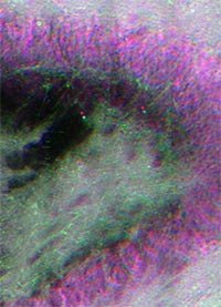
Pictured: Stimulated Raman scattering (SRS) image(s) of ‘viable’ rat DG, ~150um thick. Granule cell neurons are visible in pink (nuclei) with purple surrounds (cytoplasm). The image demonstrates that Raman imaging can be readily performed on unfixed tissue in perfusion, and the thickness of this tissue can approach hundreds of microns.
Beckman Institute for Advanced Science and Technology