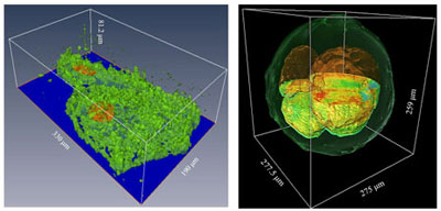
Historically, imaging techniques use florescent dyes to image cells. The problem with these dyes is that they can affect the cells themselves. It also is sometimes difficult to use them for 3D imaging because the light bounces off all the surfaces within the sample. To overcome these difficulties, the Quantitative Light Imaging Laboratory, led by Gabriel Popescu, a professor of electrical and computer engineering, has used a technique called gradient light interference microscopy or GLIM. GLIM received the 2018 Microscopy Today Method of the Year.
GLIM can be used to image 3D cellular systems such as organoids. “It can cut through multiple scattering in these structures that are hundreds of microns thick. The method allows us to visualize the tissue architecture with high resolution and contrast,” said Popescu, who is a full-time faculty member at the Beckman Institute and also affiliated with the Cancer Center at Illinois.
Two grants from the National Institutes of Health will aid in developing label-free imaging for identifying aggressive cancers as well as quantifying cell growth.
The first grant, with funding of $2 million over 4 years, will focus on developing label-free imaging for quantifying cell growth using GLIM for 3D organoids, as well as spatial light interference microscopy (SLIM) for 2D structures. A $250,000 supplement will be used to integrate GLIM in a new confocal microscope. Lab members will work with Rashid Bashir, the dean of The Grainger College of Engineering, a professor of bioengineering, and a faculty affiliate at the Beckman Institute and Carle Illinois College of Medicine.
“Bashir’s lab has been working with Mayo clinic to study microtumors for precision medicine applications. We will work with them to find out the best treatment of microtumors without using fluorescence tags,” Popescu said.
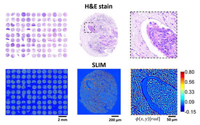
The second grant, which will provide $ 2.5 million over 5 years, will use a different label-free imaging for identifying aggressive cancers. The traditional techniques for imaging cancer biopsies involve dyes which can be used for diagnosis but are less effective in providing information on how aggressive the cancer is. The Popescu lab will partner with Carle and the University of Wisconsin-Madison to develop new intrinsic markers for patient prognosis.
Using their imaging technique, Popescu’s lab has found that as the cancer gets more aggressive, the collagen fibers surrounding it seem to provide paths for the diseased cells to metastasize. “We have found that we can make very accurate density maps of the tissues and extract information about the microenvironment of the tumor, which can tell us how fast the cancer is going to develop,” Popescu said.
The lab also plans to use computational techniques to understand the light-tissue interaction in their imaging methods. “The main limitation with label-free imaging is that sometimes you are not sure of what you are looking at; the image lacks specificity. Our recent results show that now we can recover a great deal of specificity using computation,” Popescu said. “We are set up to use data-driven approaches to extract more specific signals and believe that combining label-free imaging with artificial intelligence will lead to future breakthroughs.”
“This new funding is the result of hard work by my group members and our long-term collaborators on and off campus. I believe that our campus is very special because of how productively people work together across diverse disciplines,” Popescu said.