Breast cancer is among the top 10 leading causes of death among women worldwide. Although the treatment methods have improved, detecting and treating metastases of cancer in lymph nodes remains a challenge. Currently, doctors use methylene blue, a blue dye, to identify where the tumors aggregate in lymph nodes. However, the dye can cause skin lesions.
Members of the Pan Research Group — led by Dipanjan Pan, an associate professor of bioengineering and of materials science and engineering — are addressing this issue by developing a biocompatible nanoparticle, which will not cause any toxic side effects. Their findings have been published in “Biodegradable Biliverdin Nanoparticles for Efficient Photoacoustic Imaging” in the journal ACS Nano.

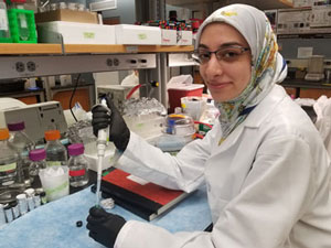
“By developing nanoparticles that are completely degradable, we hope to provide a tool that can be used for biomedical imaging and cancer therapy,” said Parinaz Fathi, the first author of the paper and a bioengineering graduate student in the Pan lab.
The lab’s biodegradable nanoparticles are made by linking together biliverdin molecules. Biliverdin is a pigment molecule present in the body. Therefore, the body is capable of breaking it down completely. Additionally, biliverdin can give a strong signal that can be easily detected.
“Our goal was to develop an agent that lights up within the body and then disappears completely after transmitting the information back,” Pan said.
In collaboration with Jefferson Chan’s lab in chemistry, the nanoparticles have been tested in breast cancer cell lines, as well as in mice. In both cases, they had no adverse effects. Chan is an associate professor of chemistry and an affiliate in biochemistry in the School of Molecular and Cellular Biology.
“We were excited to prove the complete degradability of these nanoparticles, which isn’t always possible with the currently reported nanoparticles,” Pan said.
Additionally, the particles emitted a signal, which was used to identify lymph nodes and confirm the degradation of the particles. “The inherent optical properties of biliverdin allow us to use it without needing any additional, non-degradable chemicals,” Fathi said.
"Striking the perfect balance between diagnostic performance and biocompatibility is an immense challenge that this work has successfully achieved," Chan said.
The Pan lab hopes to use these particles as vehicles for targeted cancer therapy as well as in other imaging technologies.
Pan also is associate head of Graduate Programs for the Department of Bioengineering and a faculty member at the Carle Illinois College of Medicine.
The paper “Biodegradable Biliverdin Nanoparticles for Efficient Photoacoustic Imaging” is available online:
DOI: 10.1021/acsnano.9b01201