Speakers and abstracts:
Focal frontal brain lesions predict elevated body mass index
Joachim Operskalski
Variance in adiposity and general body composition can be attributed to differences in a wide variety of biological and psychological phenomena, including hormonal appetite and satiety signaling pathways, metabolic rates, activity levels, and preferences for foods of varying nutritional quality. Neurological processes corresponding to such predictors of adiposity have been identified by neuroimaging methods, but their functional significance and the direction of causality are unclear; whether maintaining a healthy body composition truly relies on activity in specific structures in the cerebral cortex has heretofore eluded the field of human neuroscience. We thus report the findings of a large-sample human brain lesion study that helps resolve some of the ambiguity: focal damage to left-hemispheric medial prefrontal cortical region reliably predicts elevated body mass index scores among those within the unhealthy range of the body composition spectrum. Beyond the intrinsic value of mapping behavior and brain function, these findings have the potential to influence the ever-changing landscape of medical care, with possible applications to both brain injury rehabilitation and obesity prevention; given the fact that obesity is a major cause of mortality, morbidity and medical expenditure, there are clear public health implications and matters of healthcare reimbursement policy at stake.
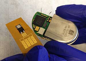
Conformal Piezoelectric Energy Harvesting From the Motion of the Heart, Lung and Diaphragm With Capacity to Operate a Cardiac Pacemaker
Canan Dagdeviren
Quantitative Ultrasound from Biophantoms to Tumors
Aiguo Han
Modern imaging plays a vital role in improving detection, classification and management of diseases such as cancer. Ultrasound is a cost-effective and safe modern imaging modality. While conventional B-mode ultrasound images display large-scale structures (greater than wavelength) of tissue, Quantitative Ultrasound (QUS), a quantitative approach, offers the capacity to quantify tissue microstructure (smaller than wavelength). A model-based approach for QUS is to develop ultrasonic scattering models that match the anatomic geometry of the tissue type under investigation. However, tissues are complex acoustic scattering media and to date there is no adequate scattering model that fits tissues. To develop appropriate models for tissue, we use a reasonable and step-wise approach. We study scattering from media of different degrees of complexity: 1) simple (low-concentration cell pellet biophantoms); 2) moderately complex (high-concentration cell pellet biophantoms); 3) significantly complex (tumors). This approach improves our understanding of acoustic scattering in biological media, and opens up opportunities to improve imaging capabilities of QUS.