Neural Correlates of Externally and Internally Originating Emotional Distraction Effects on Working Memory
Alexandru D. Iordan
Balanced emotion-cognition interactions are critical for healthy functioning. Clinical studies suggest that symptoms of impaired executive control and enhanced emotional distractibility observed in affective disorders are linked to dysfunctional interactions between a dorsal executive system and a ventral emotional system. Recent studies investigating the neural correlates of working memory interference by emotional distraction have provided evidence that interactions between these two systems can also occur transiently, in response to on-going task irrelevant emotional distracters. However, these previous investigations have used negative external distracters, such as high-arousing unpleasant pictures, and hence it is not known whether similar effects are produced by distracters with different characteristics, such as positive distracters, or distracters originating from ‘within the individual’, more similar to the distressing thoughts that occur in clinical patients. These issues have been addressed in two experiments part of an on-going investigation of the neural circuitries linking the enhancing and impairing effects of emotion on memory. Preliminary results suggest that the opposing pattern of activity in response to emotional distraction in the dorsal and ventral brain regions (decreased vs. increased) is mainly sensitive to emotional arousal rather than to emotional valence and it is similar for distracters originating in the external and internal environments.
Graphene Atomic Shrink Wrap for Nanosandwiching Biomolecules
Joshua D. Wood
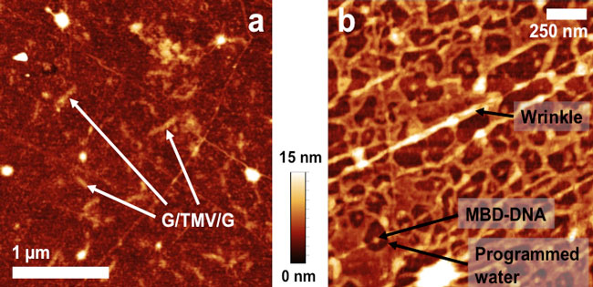
Graphene, a two-dimensional honeycomb sheet of carbon atoms, has been hotly scrutinized for its desirable electronic applications. Still, graphene has hardly been used as an atomically thin membrane. Graphene membranes are conformal to surfaces, entrapping liquids, gases, and nanoparticles. Here, we use centimeter sheets of grown graphene membranes on Cu to make graphene-biomolecule nanosandwiches. To that end, we cleanly layer graphene using a novel polymer support, poly(bisphenol A carbonate). We nanosandwich PC-transferred graphene sheets with tobacco mosaic viruses (TMV), NeutrAvidin (NA) proteins, and a complex of MBD1 protein attached to methylated DNA (MBD-DNA). The graphene layers act as atomic shrink wrap, protecting the biomolecules from environments like ultrahigh vacuum. Unusually, nanosandwiched proteins give bizarre hydration dynamics and denaturation characteristics. NA and MBD-DNA are ~5-6 nm in diameter and differ in their hydrophobic character. Nonetheless, these proteins “program” water, screening water away from the protein in a 10-fold larger radius. Nanosandwiched MBD-DNA do not change in shape when heated past the proteins’ melting point. We explain our denaturation phenomena in these nanosandwiches with molecular dynamics simulations. Our results suggest that the graphene-biomolecule heterostructures are unique preservation cells, suited for assessing atomic-scale hydration. Moreover, we show preliminary work involving these nanosandwiches in atomistic studies like scanning tunneling microscopy.
Figure: Graphene-biomolecule nanosandwiches. (a) Graphene-tobacco mosaic virus-graphene nanosandwich. (b) Graphene-MBD protein-graphene nanosandwich.
Frontoparietal traffic signals: A fast optical imaging study of preparatory dynamics in response mode switching¹
Pauline L. Baniqued
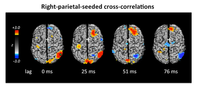
Abstract: Coordination between networks of brain regions is important for optimal cognitive performance, especially in attention demanding tasks. With the event-related optical signal (EROS; a measure of changes in optical scattering due to neuronal activity) we can characterize rapidly evolving network processes by examining the millisecond-scale temporal correlation of activity in distinct regions during the preparatory period of a response-mode switching task. Participants received a pre-cue indicating whether to respond vocally or manually. They then saw or heard the letter “L” or “R”, indicating a “left” or “right” response to be implemented with the appropriate response modality. We employed lagged cross-correlations to characterize the dynamic connectivity of preparatory processes. Our results confirmed coupling of frontal and parietal cortices, and the trial-dependent relationship of the right frontal cortex with response preparation areas. The frontal-to-modality-specific cortex cross-correlations revealed a pattern in which first irrelevant regions were deactivated and then relevant regions were activated. These results provide a window into the sub-second-scale network interactions that flexibly tune to task demands.
Figure: From publication¹.
- Baniqued, P.L., Low, K.A., Fabiani, M. & Gratton, G. (2013). Frontoparietal traffic signals: A fast optical imaging study of preparatory dynamics in response mode switching. Journal of Cognitive Neuroscience, In Press. doi:10.1162/jocn_a_00341